IMAGES IN CARDIOLOGY
Thrombi in Both Atria Detected by Cardiovascular
Tomography in a Patient with Atrial Fibrillation
Trombos en
ambas aurículas en paciente con fibrilación auricular detectados por tomografía
cardiovascular
Mariano L. Falconi1,
MTSAC, Priscila
S. Quintana1, MTSAC, Diego Pérez de Arenaza1, MTSAC
1 Cardiovascular
Imaging Section, Cardiology Service, Hospital Italiano
de Buenos Aires.
Rev Argent Cardiol 2023;91:330-331. http://dx.doi.org/10.7775.rac.v91.i4.20661
An 84-year-old female patient with a history of
hypertension, diabetes, stroke and atrial fibrillation under anticoagulant
treatment with acenocoumarol presented with severe
aortic stenosis (peak gradient 74 mmHg, mean gradient 44 mmHg, area 0.6 cm2)
and mildly depressed global left ventricular function. A cardiovascular
tomography was performed to schedule a transcatheter
aortic valve implantation (TAVI).
The study showed filling defects in early and late
phases contrast-enhanced images in the roof of the right atrium near the atrial
appendage and in the left atrial appendage, consistent with thrombi (Figures 1 and 2).
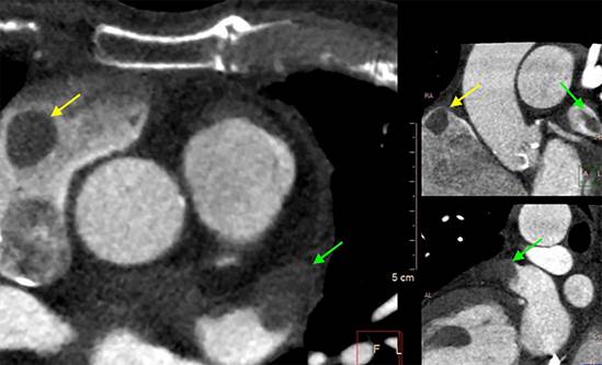
Fig.
1. Early phase contrast-enhanced images. Filling defects consistent with mass in the roof of the right
atrium (yellow arrow) and distal body of left atrial appendage (green arrow).
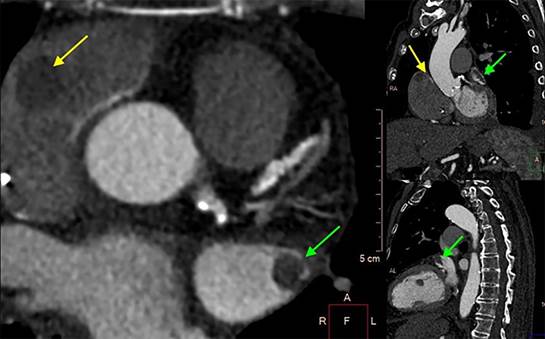
Fig.
2. Late phase contrast-enhanced images. Defect
persistence in both atria
Thrombosis of the left atrial appendage is frequent in
atrial fibrillation, and its prevalence (up to 8% with anticoagulant therapy
and 5-27% without) depends largely on the studied population. However,
thrombosis of the right atrial appendage is rare (estimated at 0.6-0.75%), and
its incidence is lower due to its anatomy (more open and with less potential
for blood stasis –“remora phenomenon”– than the left one) and to a search
performed less systematically as well as difficulties in evaluating its
structure.(1) However, autopsy studies suggest that the prevalence
of thrombosis in both atria is similar.(2)
Cardiovascular tomography is an excellent tool to
evaluate atrial thrombosis, which is observed as a defined structure with clear
borders generating filling defects both in early and late phases after the
contrast agent is administered (it distinguishes from the remora phenomenon in
which defects in early phases are corrected in late phases). It has very high
sensitivity and specificity for the detection of thrombi in the left atrium.(3) However, in some cases, it is difficult to achieve an
adequate contrast in the right chambers, mainly the right atrial appendage, by
using imaging to evaluate “left structures” (coronary arteries, pulmonary
veins, valves); thus, right atrial thrombosis may be undetected. The low signal
in Hounsfield units on non-contrast-enhanced tomography or the Hounsfield units ratio between thrombus and the aorta using dual-source
equipment (4) may be helpful in identifying atrial thrombosis by
cardiovascular tomography.
Conflicts of interest
None declared (See authors’ conflicts
of interest forms on the website).
 https://creativecommons.org/licenses/by-nc-sa/4.0/
©Revista Argentina
de Cardiología
1.
García-Fernández MÁ, Cresti A. Right Atrial Appendage Thrombus: “Can Be Found
if You Look for It.” JACC Case Rep. 2023;5:101702.
https://doi.org/10.1016/j.jaccas.2022.101702
2.
Richardson AC, Omar M, Velarde G, Missov E, Percy R, Sattiraju S. Right Atrial
Appendage Thrombus in Atrial Fibrillation: A Case Report and Review of the
Literature. J Investig Med High Impact Case Rep. 2021;9:23247096211010050. https://doi.org/10.1177/23247096211010048
3. Pathan
F, Hecht H, Narula J, Marwick TH. Roles of Transesophageal Echocardiography and
Cardiac Computed Tomography for Evaluation of Left Atrial Thrombus and
Associated Pathology: A Review and Critical Analysis. JACC Cardiovasc Imaging. 2018;11:616-27. https://doi.org/10.1016/j.jcmg.2017.12.019
4. Li W,
Liu M, Yu F, Zhu W, Yu X, Guo X, Yang Q. Detection of left atrial appendage
thrombus by dual-energy computed tomography-derived imaging biomarkers in
patients with atrial fibrillation. Front Cardiovasc
Med. 202222;9:809688. https://doi.org/10.3389/fcvm.2022.809688