Alejandro Moreno1, Alfredo Matías Rodríguez Granillo1 MTSAC, Ricardo Pérez de la Hoz 1 MTSAC
1 Sanatorio Otamendi
Address for reprints: Alejandro Moreno. Sanatorio Otamendi. E-mail: morenoa@otamendi.com.ar
ARGENT J CARDIOL 2023;91:343-348.http://dx.doi.org/10.7775/rac.es.v91.i4.20669
ABSTRACT
Ischemic heart disease is the most common cause of heart failure, with a high incidence of heart failure despite early revascularization
and neurohormonal modulation.
In the acute myocardial infarction setting, necrotized cardiomyocytes induce
activation of the innate immune system, increasing the levels of inflammatory cells to help remove dead cells and initiate a
corrective response, which allows for proper scar tissue formation.
A prolonged or expanded inflammatory response after infarction contributes to adverse ventricular remodeling and development
of heart failure.
Understanding the inflammatory mechanisms that emerge as a result of myocardial infarction and their impact on adverse
remodeling that leads to an increased.
Key words: Acute Myocardial Infarction - Inflammation - Ventricular Remodeling
RESUMEN
La cardiopatía isquémica es la causa más frecuente de insuficiencia cardíaca, con una alta incidencia de esta a pesar de la revascularización precoz y la modulación neurohormonal.
En el contexto del infarto agudo de miocardio los cardiomiocitos necrosados inducen la activación del sistema inmune innato, con aumento de la concentración de células inflamatorias que ayudan a eliminar las células muertas, e iniciar una respuesta correctiva que permite la formación adecuada de tejido cicatrizal.
La prolongación o expansión de la respuesta inflamatoria posterior al infarto contribuye al remodelado adverso ventricular
y al desarrollo de insuficiencia cardíaca.
Entender los mecanismos inflamatorios que se desarrollan producto del infarto, y su impacto en el remodelado adverso que
aumenta el número de eventos cardiovasculares mayores, permite comprender a la inflamación como un objetivo terapéutico.
Received: 07/07/2023
Accepted: 09/19/2023
Palabras clave: Infarto agudo de miocardio - Inflamación - Remodelado ventricular
INTRODUCTION
ST-elevation myocardial infarction (STEMI) is a con- dition with a high global incidence and entails an increased risk of heart failure (HF) and mortality.
In the US, there is an estimated annual incidence of 605 000 new acute myocardial infarction (AMI) cases and 200 000 recurrent events. (1)
In our country, the na- tional registry of ST-elevation myocardial infarction (ARGEN-IAM-ST) estimates an incidence of 120 in- farctions per 100 000 inhabitants. (2)
After a STEMI, early and successful myocardial reperfusion thanks to thrombolytic therapy or pri- mary percutaneous coronary intervention (PCI) is the most effective strategy to reduce the extent of infarc- tion and to improve clinical outcomes.
However, the blood flow restoration process for an ischemic myo- cardium may induce lesions.
This condition, known as myocardial reperfusion damage, may ironically reduce its beneficial effects.
The lesion results in death of formerly viable cardiomyocytes immediately before reperfusion, thus increasing the extent of infarction.(3,4)
This lesion is caused by several mechanisms, such as the accumulation of neutrophils and the increase of oxidative stress due to production of reactive oxygen species (ROS), (5) which leads to sudden in- crease of intracellular calcium secondary to damage of the sarcoplasmic membrane and sarcoplasmic reticu- lum dysfunction.(4)
Studies in animal models of AMI suggest that fatal reperfusion lesions represent up to 50% of the final extent of infarction. (3)
Despite progress made in treatment of coronary disease and specifically AMI in the last two decades, it continues to be the most common cause of heart fail- ure (HF). (6)
HF after discharge in patients with AMI is very common.
It is diagnosed in about 13% of patients within 30 days, and in 20-30% of patients within a year.
HF incidence after AMI discharge is higher in the first few months, then reduced, and maintained at an annual rate of 1.3-2.2%.(7)
THE ROLE OF INFLAMMATION
There are different risk factors and mechanisms lead- ing to HF in different moments.
Among them, we can cite age, female sex, multivessel disease, previous AMI, hypertension, atrial fibrillation, diabetes, chronic kidney disease, and preconditioning.
There is increasing- ly more evidence that a prolonged or expanded inflam- matory response after AMI significantly contributes to left ventricular (LV) remodeling and HF. (8)
Inflammatory response quantification methods show prom- ising HF predictive results.
The C-reactive protein (CRP) levels anticipate a risk of adverse events after AMI, including HF.(9)
The proportion of neutrophils and lymphocytes, indicating systemic inflammation, predicted major adverse cardiovascular events (MACE) and HF in a meta-analysis of 14 studies.(10)
Cytokines are strategic inflammation regulators.
In a study of 4939 patients with acute coronary syndrome, pro-inflammatory cytokine interleukin-6 (IL-6) levels were independent MACE and HF predictors(11)
IL- 32 is a pro-inflammatory cytokine inducing the release of other inflammatory cytokines, such as the tumor necrosis factor-α (TNF-α), IL-1β, IL-6, IL-8, and IL-18. Xuan et al. showed that IL-32 is an independent pre- dictor of cardiac death and HF in patients following AMI.(12)
Emergence of acute myocardial ischemia in the AMI setting induces an initial pro-inflammatory response aimed at removing any remaining necrotic cells from the AMI area by means of various processes, including complement cascade activation, production of reactive oxygen species (ROS), and damage-associated molecular patterns (DAMP), acting as ligands for pattern recognition receptors (PRR), such as Toll-like receptors (TLR), dendritic cells, and inflammasomes
The latter release several pro-inflammatory mediators (interleukins and chemokines) inducing inflamatory cell recruitment (neutrophils, lymphocytes, monocytes, and macrophages) in the AMI area and increasing the pro-inflammatory response.
Since they are targeted to the borderline viable infarction area, infiltrated leukocytes may induce cardiomyocytes death, thus extending the ischemic lesion beyond the original infarction area. (Figure 1) (13)
Myocardial reperfusion onset following coronary angioplasty exacerbates this pro-inflammatory response and contributes to the death of cardiomyocytes and myocardial damage typical of a "myocardial reperfusion lesion" occurring from 6 to 24 h after reperfusion. (14)
The initial pro-inflammatory response is followed by an anti-inflammatory repair phase, which enables scar tissue formation that avoids heart rupture. The anti-inflammatory repair phase (days 4 to 7) follow- ing AMI is triggered by initial pro-inflammatory re- sponse suppression, resolution, and control.(Figure 2) (13)
This is driven by inhibitory pathways activation, suppressing inflammation and dynamic changes in the function of infiltrating leukocytes in the AMI area.
Lymphocytes play a role in this phase; apoptosis and removal from the infarction area are a hallmark of resolved inflammation and repair phase
This is an active process that requires recruitment of inhibitory pathways cascades resulting in anti-inflammatory M2 macrophage polarization, and both anti-inflammatory and profibrotic cytokine secretion, which suppress inflammation and promote tissue repair thanks to the action of fibroblasts.(15-17)
INFLAMMATION AND REMODELING
Following AMI, the LV undergoes geometric and func- tional changes, with hypertrophy of non-infarction segments and dilation/narrowing of infarction seg- ments, a process known as adverse remodeling and associated with poor clinical outcomes. According to a systematic review of 37 studies, the most common parameters when defining ventricular remodeling are 20% increase in left ventricular end-diastolic volume (LVEDV) and/or 15% increase in left ventricular end-systolic volume (LVESV). (18)
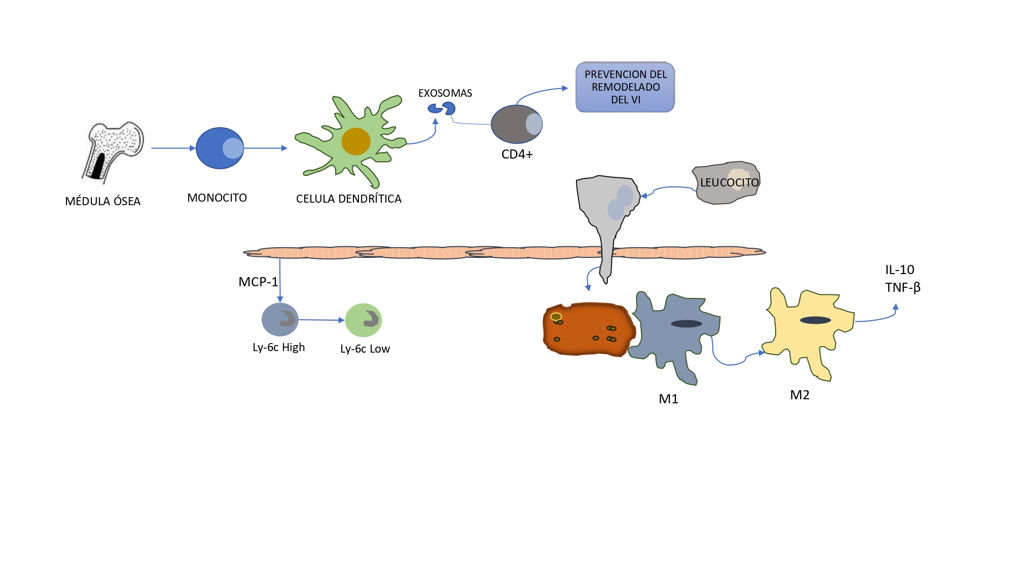
Fig. 1. DAMP-induced pro-inflammatory response.
After an AMI, release of molecular patterns associated with damage or DAMP (such as ATP, mtDNA, RNA, and HMBGB1) induces a pro-inflammatory response that mediates cardiomyocytes death by means of Toll-like receptors (TLR) and leukocyte recruitment in the infarction area, cytokine release, mitochondrial dysfunction (calcium overload and ROS production), as well as inflammasome formation. Based on Ong SB et al. Pharmacol Ther. 2018;186:73-87

Fig. 2. The anti-inflammatory repair phase following acute myocardial infarction.
After the AMI pro-inflammatory response, the anti-inflammatory repair phase leads to inflammation resolution.
The bone marrow and circulating monocytes are distinguished in dendritic cells that prevent LV remodeling through CD4 + leukocytes exosome activation.
Apoptotic neutrophil expression induces M2 macrophage polarization and secretion of anti-inflammatory and profibrotic cytokines, such as IL-10 and TGF-β, which suppress inflammation and promote tissue repair.
A change from high pro-inflammatory Ly6c monocytes and M1 macrophages located in the AMI area in response to increased MCP-1 myocardial expression during the initial pro-inflammatory phase to low anti-inflammatory Ly6C, monocytes, and M2 macrophages. Based on Ong SB et al. Pharmacol Ther. 2018;186:73-87
An excessive and persistent pro-inflammatory response after AMI can aggravate LV adverse remodeling by increasing inflammatory cytokine expression.
These induce cardiomyocyte apoptosis, increase the extracellular matrix deposit, and depress contractility, which results in a more rigid ventricle, with larger diastolic dysfunction and fibroblast activation in the remote myocardium that may expand fibrosis in viable tissue. (19)
Experimental and clinical studies establish a relationship between interleukin expression and LV volumes. (20-22)
A first-line method to determine the incidence and extent of ventricular remodeling is 2D ultrasound.
In an ultrasound sub-study of the VALIANT trial (valsartan versus captopril or their combination in AMI complicated by systolic dysfunction or HF), the initial LV ejection fraction (LVEF), LVEDV and LVESV were independent predictors of the combined primary end-point of death or hospitalization due to heart failure(23)
Also, as compared to patients with no evidence of LV remodeling, patients with any of the LV remodeling patterns after myocardial infarction have a higher risk of cardiovascular death, AMI, HF, stroke, and cardiac arrest with reanimation.(24)
Therefore, while LV volumes continue to be strong cardiovascular prognosis indicators, the specific LV remodeling pattern contains additional information.
Cardiac magnetic resonance (CMR) imaging remains the gold standard to evaluate cardiac anatomy and function.
In addition to providing accurate meas- urement of ventricular volumes, it makes it possible to define the AMI extent in grams.
The latter is a significant predictor of adverse ventricular remodeling.
The larger the extent of the infarction, the higher the subsequent increase in LVEDV and LVESV.
The AMI extent is a better adverse remodeling predictor than final LVEF. (25)
A CMR measures native T1 (T1 mapping) in the heart with no gadolinium requirement, though usually required to outline infarction tissue.
The T1 is affected by water content, macromolecular binding, and cell composition.
Tissue water content increases as a result of ischemia, and this leads to longer T1 times and a more marked myocardial damage biomarker in localized myocardial regions. (26)
Myocardial native T1 is increased by inflammatory cell infiltration, as revealed by histopathology in patients undergoing heart transplantation with acute rejection features.(27)
Native T1 in the remote myocardium in STEMI survivors is independently linked to systemic inflammation and the extent of infarction.
Changes in the number of circulating monocytes following myocar- dial infarction is independently associated with native T1 in the remote area and subsequent LV remodeling after 6 months, supportive of a mechanical association between inflammation and adverse remodeling.
This is also related to changes in LVEDV after 6 months and independently associated with adverse cardiac events after discharge, including all-cause death and hospitalization due to HF during longer term follow-up. (28)
THERAPEUTIC CONSIDERATIONS
Given the harmful effects of an excessive and persis- tent pro-inflammatory response to AMI, and the ben- eficial effects of the repair anti-inflammatory phase that follows, a potential therapeutic strategy to limit the extent of AMI and prevent LV adverse remodeling is to mitigate the pro-inflammatory response and up-regulate the subsequent anti-inflammatory repair response.
This provides an additional therapeutic target to prevent HF after AMI.
Several therapy approaches targeted to addressing the pro-inflammatory response after AMI have been investigated, and many of them unfortunately fail to show any benefit from a reduced extent or any improved clinical outcomes.
The first experimental studies using non-specific anti-inflammatory agents, such as corticosteroids and nonsteroidal anti-inflammatory drugs (NSAIDs), showed reduced AMI extent in dogs.(29)
This was associated with adverse events, such as impaired myo- cardial repair, and cardiac narrowing and rupture, and, therefore, use of these drugs is avoided. (30)
The complement cascade is another pathway considered for AMI inflammation modulation.
In experimental studies, inhibition of this cascade reduced the final AMI extent.
However, in clinical trials, using pexelizumab, a C5 complement antibody, failed to show any clinical benefits or extent of infarction. (31)
Inflammatory cytokines are interesting therapeutic targets due to their pleiotropic effects on immune response
In the VCU-ART 3 trial, anakinra, an IL-1 inhibiting antibody, administered for 14 days following AMI significantly reduced CRP levels, death incidence, or de novo HF and HF hospitalization.(32)
An analysis of VCU-ART1, VCU-ART2 and VCU-ART3 trials is consistent with the results of the latter. (33)
CANTOS was another trial aimed to inhibit interleukin expression, in this case, IL-1 , by means of a monoclonal antibody, canakinumab.
SC administration of this drug to patients with previous AMI and elevated CRP prevents recurrent cardiovascular events (cardiovascular death, non-fatal AMI, non-fatal stroke). (34)
Later, Everett et al. found that reduced HF by canakinumab and the HF hospitalization and death compound are dose related. (35)
Metoprolol reduces neutrophil myocardial infiltration by inhibiting the β1-adrenergic receptor selective antagonist (ADRB1) in animals with provoked AMI. (36)
In the METOCARD-CNIC trial, IV administration of this betablocker reduced the extent of infarction and increased LVEF on CMR imaging versus placebo in AMI patients. (37)
Sodium-glucose linked transporter type 2 (SGLT2) inhibitors have a pleiotropic effect, and their anti-inflammatory effect is studied in experimental and clinical trials.(38)
They have shown to mitigate macrophage infiltration, particularly, M1 polarization, thus increasing the polarity of M2 involved in the anti-inflammatory phase; therefore, it is clear that SGLT2 inhibitors take part in inflammation modulation and tissue repair.(39)
In addition, they reduce the TNF-α factor expression, monocyte chemoattractant protein-1 (MCP-1), platelet adhesion molecules, IL-6, and IL-1β.
In clinical trials of diabetic and chronic coronary disease patients, treatment with SGLT2 inhibitors significantly reduces CRP levels in blood, inflammatory cytokines (IL-6), and TNF-α. (40)
As a result, treatment with this drug may improve cardio-vascular outcomes thanks to its benefits for inflammation modulation.
In terms of ventricular remodeling, in the SUGAR-DM-HF trial, using empagliflozin in HF patients with reduced LVEF and diabetes was associated with larger inverse remodeling measured by CMR.(41)
The effects of early administration of SGLT2 inhibitors in patients with recent AMI are still not fully known.
We have recently become aware of the results of the EMMY study.
In this trial, the administration of 10 mg orally of empagliflozin within 72 hours of angioplasty in patients with AMI was significantly associated with a decrease of NT-proBNP, higher LVEF, decreased LVEDV and LVESV than the placebo group.(42)
Two other ongoing studies are evaluating the safety and efficacy of SGLT2 inhibitors in patients with AMI: EMPACT-MI (NCT04509674) and DAPA-MI (NCT04564742).
CONCLUSION
LV remodeling is associated with poor outcomes in AMI patients. Therefore, it is very important to evalu- ate adverse ventricular remodeling, since reversing, stopping or at least delaying it is an essential goal of HF therapy.
Inflammation plays a crucial role in adverse ven- tricular remolding after AMI.
Exposure to underlying mechanisms identifies a series of therapeutic targets to reduce AMI extent and to prevent adverse remodeling.
It is based on pro- inflammatory phase suppression and positive regula- tion of the anti-inflammatory response
It is proposed as a complement to known treatment with large ben- efits for AMI patients, such as early and complete re- vascularization, and treatment with neurohormonal antagonists.
https://creativecommons.org/licenses/by-nc-sa/4.0/

©Revista Argentina de Cardiología
REFERENCES
- Virani SS, Alonso A, Benjamin EJ, Bittencourt MS, Callaway CW, Carson AP, et al. American Heart Association Council on Epidemiology and Prevention Statistics Committee and Stroke Statistics Subcommittee. Heart Disease and Stroke Statistics-2020 Update: A Report From the American Heart Association. Circulation. 2020;141:e139- e596. https://doi.org/10.1161/CIR.0000000000000757
- Cohen Arazi H, Zapata G, Marturano MP, De la Vega MB, Pellizón OA, D Imperio H, y cols. Angioplastia primaria en Argentina. Registron ARGEN-IAM-ST (Relevamiento Nacional del infarto agudo de miocardio con elevación del segmento ST) [Primary angioplasty in Argentina. Results from ARGEN-IAM-ST registry]. Medicina (B Aires). 2019;79:251-6. Spanish.
- Yellon DM, Hausenloy DJ. Myocardial reperfusion injury. N Engl J Med. 2007;357:1121-35. https://doi.org/10.1056/NEJMra071667
- Zweier JL, Talukder MA. The role of oxidants and free radicals in reperfusion injury. Cardiovasc Res. 2006;70:181-90. https://doi.org/10.1016/j.cardiores.2006.02.025
- Litt MR, Jeremy RW, Weisman HF, Winkelstein JA, Becker LC. Neutrophil depletion limited to reperfusion reduces myocardial infarct size after 90 minutes of ischemia. Evidence for neutrophilmediated reperfusion injury. Circulation. 1989;80:1816-27. https://doi.org/10.1161/01.cir.80.6.1816
- de Jaegere PPT, de Weger A, den Heijer P, Verkroost M, Baan J, de Kroon T, et al.Treatment decision for transcatheter aortic valve implantation: the role of the heart team : Position statement paper of the Dutch Working Group of Transcatheter Heart Interventions. Neth Heart J. 2020;28:229-39.https://doi.org/10.1007/s12471-020-01367-4
- Fried LP, Tangen CM, Walston J, Newman AB, Hirsch C, Gottdiener J, et al. Frailty in older adults: evidence for a phenotype. J Gerontol A Biol Sci Med Sci. 2001;56:M146-56. https://doi.org/10.1093/gerona/56.3.M146
- Otto CM, Baumgartner H. Updated 2017 European and American guidelines for prosthesis type and implantation mode in severe aortic stenosis. Heart. 2018;104:710-3. https://doi.org/10.1136/heartjnl-2017-312487
- Dubois C, Coosemans M, Rega F, Poortmans G, Belmans A, Adriaenssens T, et al. Prospective evaluation of clinical outcomes in all-comer high-risk patients with aortic valve stenosis undergoing medical treatment, transcatheter or surgical aortic valve implantation following heart team assessment. Interact Cardiovasc Thorac Surg. 2013;17:492-500. https://doi.org/10.1093/icvts/ivt228
- Costa C, Teles RC, Brito J, Neves JP, Gabriel HM, Abecassis M, et al. Advantages of a prospective multidisciplinary approach in transcatheter aortic valve implantation: Eight years of experience. Rev Port Cardiol. 2017; 36:809-18. https://doi.org/10.1016/j.repc.2016.11.015
- Iglesias D, Salinas P, Moreno R, García-Blas S, Calvo L, Jiménez- Valero S, et al. Prognostic impact of decisions taken by the heart team in patients evaluated for transcatheter aortic valve implantation. Rev Port Cardiol 2015;34:587-95. https://doi.org/10.1016/j.repc.2015.03.013
- Bouleti C, Chauvet M, Franchineau G, Himbert D, Iung B, Alos B, et al. The impact of the development of transcatheter aortic valve implantation on the management of severe aortic stenosis in high-risk patients: treatment strategies and outcome. Eur J Cardiothorac Surg. 2017;51:80-8. https://doi.org/10.1093/ejcts/ezw211
- Antonides C, Mack M, Kappetein A. Approaches to the Role of The Heart Team in Therapeutic Decision Making for Heart Valve Disease. Structural Heart 2017;1:249-55. https://doi.org/10.1080/24748706.2017.1380377
- Giordano A, Biondi-Zoccai G, Frati G, Bartorelli AL. Management of Structural Heart Disease and Acute Coronary Syndromes in the COVID-19 Pandemic. Curr Atheroscler Rep. 2020;22:29-35. https://doi.org/10.1007/s11883-020-00849-5
- Burzotta F, Graziani F, Trani C, Aurigemma C, Bruno P, Lombardo A, et al. Clinical Impact of Heart Team Decisions for Patients with Complex Valvular Heart Disease: A Large, Single-Center Experience. J Am Heart Assoc. 2022;11:e024404. https://doi.org/10.1161/JAHA.121.024404
- Khan S, Shi W, Kaneko T, Baron SJ. The Evolving Role of the Multidisciplinary Heart Team in Aortic Stenosis. US Cardiology Review 2022;16:e19. https://doi.org/10.15420/usc.2022.04
- Leon MB, Smith CR, Mack M, Miller DC, Moses JW, Svensson LG, et al and PARTNER Trial Investigators. Transcatheter aortic-valve implantation for aortic stenosis in patients who cannot undergo surgery. N Engl J Med. 2010;363:1597-607. https://doi.org/10.1056/NEJMoa1008232
- Reardon MJ, Van Mieghem NM, Popma JJ, Kleiman NS, Søndergaard L, Mumtaz M, et al. Surgical or Transcatheter Aortic- Valve Replacement in Intermediate-Risk Patients N Engl J Med 2017;376:1321-31. https://doi.org/10.1056/NEJMoa1700456
- Garmendia CM, Seropian IM, Chiabrando JG, Medina de Chazal H, Cal M, Kotowicz V, y cols. Prevalencia, predictores e impacto clínico de la rehospitalización en pacientes con estenosis valvular aórtica valorados por un Heart Team. Rev Argent Cardiol 2021;89:501-6. http://dx.doi.org/10.7775/rac.es.v89.i6.20459
- Adams HSL, Ashokkumar S, Newcomb A, MacIsaac AI, Whitbourn RJ, Palmer S. Contemporary review of severe aortic stenosis. Intern Med J. 2019;49:297-305. http://dx.doi.org/10.1111/imj.14071
- Youn S, Wong SA, Chrystoja C, Tomlinson G, Wijeysundera HC, Bell CM, et al. Bias estimation in study design: a meta-epidemiological analysis of transcatheter versus surgical aortic valve replacement. BMC Surg. 2021;21:285. http://dx.doi.org/10.1186/s12893-021-01278-0
- Ørn S, Ueland T, Manhenke C, Sandanger Ø, Godang K, Yndestad A, et al. Increased interleukin-1β levels are associated with left ventricular hypertrophy and remodelling following acute ST segment elevation myocardial infarction treated by primary percutaneous coronary intervention. J Intern Med. 2012;272:267-76.Hppts://doi.org/10.1111/j.1365-2796.2012.02517.x
- Solomon SD, Skali H, Anavekar NS, Bourgoun M, Barvik S, Ghali JK, et al. Changes in ventricular size and function in patients treated with valsartan, captopril, or both after myocardial infarction. Circulation. 2005;111:3411-9.https://doi.org/10.1161/CIRCULATIONAHA.104.508093
- Verma A, Meris A, Skali H, Ghali JK, Arnold JM, Bourgoun M, et al. Prognostic implications of left ventricular mass and geometry following myocardial infarction: the VALIANT (VALsartan In Acute myocardial iNfarcTion) Echocardiographic Study. JACC Cardiovasc Imaging. 2008;1:582-91. https://doi.org/10.1016/j.jcmg.2008.05.012
- Wu E, Ortiz JT, Tejedor P, Lee DC, Bucciarelli-Ducci C, Kansal P, et al. Infarct size by contrast enhanced cardiac magnetic resonance is a stronger predictor of outcomes than left ventricular ejection fraction or end-systolic volume index: prospective cohort study. Heart. 2008;94:730-6. https://doi.org/10.1136/hrt.2007.122622
- Mathur-De Vré R. Biomedical implications of the relaxation behaviour of water related to NMR imaging. Br J Radiol. 1984;57:955-76. https://doi.org/10.1259/0007-1285-57-683-955
- Miller CA, Naish JH, Shaw SM, Yonan N, Williams SG, Clark D, et al. Multiparametric cardiovascular magnetic resonance surveillance of acute cardiac allograft rejection and characterisation of transplantation-associated myocardial injury: a pilot study. J Cardiovasc Magn Reson. 2014;16:52. https://doi.org/10.1186/s12968-014-0052-6
- Carrick D, Haig C, Rauhalammi S, Ahmed N, Mordi I, McEntegart M, et al. Pathophysiology of LV Remodeling in Survivors of STEMI: Inflammation, Remote Myocardium, and Prognosis. JACC Cardiovasc Imaging. 2015;8:779-89. https://doi.org/10.1016/j.jcmg.2015.03.007
- Kirlin PC, Romson JL, Pitt B, Abrams GD, Schork MA, Lucchesi BR, et al. Ibuprofen-mediated infarct size reduction: effects on regional myocardial function in canine myocardial infarction in canine myocardial infarction. Am J Cardiol. 1982;50:849-56. https://doi.org/10.1016/0002-9149(82)91244-9
- Hammerman H, Kloner RA, Hale S, Schoen FJ, Braunwald E. Dose-dependent effects of short-term methylprednisolone on myocardial infarct extent, scar formation, and ventricular function. Circulation. 1983;68:446-52. https://doi.org/10.1161/01.cir.68.2.446
- Mahaffey KW, Granger CB, Nicolau JC, Ruzyllo W, Weaver WD, Theroux P,et al. COMPLY Investigators. Effect of pexelizumab, an anti-C5 complement antibody, as adjunctive therapy to fibrinolysis in acute myocardial infarction: the COMPlement inhibition in myocardial infarction treated with thromboLYtics (COMPLY) trial. Circulation. 2003;108:1176-83. https://doi.org/10.1161/01.CIR.0000087404.53661.F8
- Abbate A, Trankle CR, Buckley LF, Lipinski MJ, Appleton D, Kadariya D, et al. Interleukin-1 Blockade Inhibits the Acute Inflammatory Response in Patients With ST-Segment-Elevation Myocardial Infarction. J Am Heart Assoc. 2020;9:e014941. https://doi.org/10.1161/JAHA.119.014941
- Abbate A, Wohlford GF, Del Buono MG, Chiabrando JG, Markley R, Turlington J, et al. Interleukin-1 blockade with anakinra and heart failure following ST-segment elevation myocardial infarction: results from a pooled analysis of the VCUART clinical trials. Eur Heart J Cardiovasc Pharmacother. 2022;8:503-10. https://doi.org/10.1093/ehjcvp/pvab075
- Ridker PM, Everett BM, Thuren T, MacFadyen JG, Chang WH, Ballantyne C, et al; CANTOS Trial Group. Antiinflammatory Therapy with Canakinumab for Atherosclerotic Disease. N Engl J Med. 2017;377:1119-31. https://doi.org/10.1056/NEJMoa1707914
- Everett BM, Cornel JH, Lainscak M, Anker SD, Abbate A, Thuren T, et al. Anti-Inflammatory Therapy With Canakinumab for the Prevention of Hospitalization for Heart Failure. Circulation.2019 Mar;139:1289-99.https://doi.org/10.1161/CIRCULATIONAHA.118.038010
- García-Prieto J, Villena-Gutiérrez R, Gómez M, Bernardo E, Pun-García A, García-Lunar I, et al. Neutrophil stunning by metoprolol reduces infarct size. Nat Commun. 2017;8:14780. https://doi.org/10.1038/ncomms1478.
- Ibanez B, Macaya C, Sánchez-Brunete V, Pizarro G, Fernández- Friera L, Mateos A, et al. Effect of early metoprolol on infarct size in ST-segment-elevation myocardial infarction patients undergoing primary percutaneous coronary intervention: the Effect of Metoprolol in Cardioprotection During an Acute Myocardial Infarction (METOCARD-CNIC) trial. Circulation. 2013;128:1495-503. https://doi.org/10.1161/CIRCULATIONAHA.113.003653
- Nakatsu Y, Kokubo H, Bumdelger B, Yoshizumi M, Yamamotoya T, Matsunaga Y, et al. The SGLT2 Inhibitor Luseogliflozin Rapidly Normalizes Aortic mRNA Levels of Inflammation-Related but Not Lipid-Metabolism-Related Genes and Suppresses Atherosclerosis in Diabetic ApoE KO Mice. Int J Mol Sci. 2017;18:1704. https://doi.org/10.3390/ijms18081704
- Xu L, Ota T. Emerging roles of SGLT2 inhibitors in obesity and insulin resistance: Focus on fat browning and macrophage polarization. Adipocyte. 2018;7:121-8. https://doi.org/10.1080/21623945.2017.1413516
- Oikonomou E, Xenou M, Zakynthinos GE, Tsaplaris P, Lampsas S, Bletsa E, et al. Novel Approaches to the Management of Diabetes Mellitus in Patients with Coronary Artery Disease. Curr Pharm Des. 2023;29:1844-62.https://doi.org/10.2174/1381612829666230703161058
- Lee MMY, Brooksbank KJM, Wetherall K, Mangion K, Roditi G, Campbell RT, et al. Effect of Empagliflozin on Left Ventricular Volumes in Patients With Type 2 Diabetes, or Prediabetes, and Heart Failure With Reduced Ejection Fraction (SUGAR-DM-HF). Circulation. 2021;143:516-25. https://do.org/10.1161/CIRCULATIONAHA.120.052186
- von Lewinski D, Kolesnik E, Tripolt NJ, Pferschy PN, Benedikt M, Wallner M, et al. Empagliflozin in acute myocardial infarction: the EMMY trial. Eur Heart J. 2022;43:4421-32. https://doi.org/10.1093/eurheartj/ehac494