

Pathophysiological Basis
Cardiovascular (CVD) and metabolic diseases are the leading cause of mortality in Western societies. Metabolic syndrome (MS) represents the combination of several risk factors, including obesity, dyslipidemia, hyperglycemia, oral glucose intolerance and hypertension, and contributes to the development of CVD and type 2 diabetes mellitus (T2DM). (1)
Feeding rats with high fat and carbohydrate diets—which mirror the Western population diet—induces the development of various components of MS. Rats fed a high-fat diet supplemented with fructose (10%) in the drinking water for 8–9 weeks exhibit increased triglyceride and cholesterol levels, as well as insulin resistance, visceral adiposity and blood pressure, in addition to ventricular hypertrophy, myocardial fibrosis, alterations in stroke volume, and remodeling of the coronary arteries. These pathophysiological changes involve increased oxidative, inflammatory and fibrotic processes, as well as mitochondrial dysfunction. (2)
On the other hand, overweight and obesity are frequently associated with mineral and vitamin deficiencies. (3) Several meta-analyses have reported low plasma zinc concentrations, hyperzincuria and redistribution of this mineral in diabetic and obese patients. (4) Furthermore, it has been shown that chronic low zinc intake is associated with reduced insulin secretion and/or tissue resistance to insulin, increased blood glucose levels, and alterations in lipid metabolism. (5) We have previously demonstrated that moderate zinc deficiency during prenatal and early postnatal development leads to renal, cardiovascular, and metabolic abnormalities in adult rats. In our experimental studies, male rats subjected to this deficiency exhibited lower birth weight, reduced renal and cardiac tissue weights, increased blood pressure, cardiorenovascular dysfunction, related to inadequate activity of the nitric oxide (NO) and renal and cardiovascular renin-angiotensin systems, as well as increased renal oxidative stress. They also exhibited hypertriglyceridemia, hyperglycemia, insulin resistance, hypertrophy, and increased oxidative state of the retroperitoneal adipose tissue. Females appear to be less sensitive to this nutritional injury. (6) In addition, zinc deficiency during fetal and postnatal life was shown to exacerbate the metabolic and retroperitoneal adipose tissue alterations induced by a high-fat diet during growth. (7)
There is an urgent need to identify new therapeutic strategies aimed at reducing the cardiovascular morbidity and mortality associated with MS. Selective sodium-glucose cotransporter 2 inhibitors (SGLT2i), such as empagliflozin, dapagliflozin (DAPA), and canagliflozin, belong to a new class of drugs approved for use as antihyperglycemic therapy. SGLT2 cotransporters transport glucose and sodium in a 1:1 ratio and are located on the apical membrane of proximal tubules S1 and S2 segments. In healthy individuals, 90% of filtered glucose is reabsorbed in the renal proximal tubule by SGLT2, which has high affinity and capacity. The remaining 10% of filtered glucose is reabsorbed by SGLT1 transporters, which have higher affinity but lower capacity than the SGLT2 isoform. SGLT1 is located on the luminal membrane of the proximal tubule S2 and S3 segments, with a glucose-sodium coupling ratio of 1:2. When SGLT2 is blocked, SGLT1 can account for up to 50% of glucose reabsorption, since two sodium ions must be taken up for every glucose molecule absorbed. On the basolateral side, glucose leaves the intercellular space by passive diffusion through the GLUT1 and GLUT2 transporters, while sodium is expelled into the interstitium by the sodium-potassium ATPase pump. Moreover, SGLT2 transporters are physically and functionally coupled to the sodium-proton exchanger 3 (NHE3). Inhibition of SGLT2 reduces NHE3 activity, further decreasing proximal sodium reabsorption and contributing to natriuresis. (1, 8-10)
The filtered glucose load increases as plasma glucose concentration rises, with a maximum renal reabsorption capacity of approximately 375 mg/min in a healthy individual. This typically occurs at blood glucose levels between 180 and 215 mg/dL, whereas higher blood glucose levels lead to glucosuria. In people with T2DM, the threshold at which glucose appears in urine, as well as the maximum capacity for renal tubular reabsorption, increase significantly due to SGLT2 overexpression. Therefore, SGLT2i or gliflozins reduce glucose and sodium reabsorption in the proximal tubule and cause osmotic diuresis by inducing glucosuria and natriuresis. Consequently, they lower glycated hemoglobin and postprandial blood glucose levels through an insulin-independent mechanism. Their effects are not affected by impaired pancreatic islet β-cell function or by reduced insulin sensitivity. Nor are they associated with a risk of hypoglycemia, even in non-diabetic individuals. (8-10)
By lowering blood glucose levels, SGLT2i reduce cellular damage caused by advanced glycation end products, oxidative stress, inflammation, and apoptosis. They also promote the oxidation of glucose and long-chain fatty acids, regardless of insulin levels, with a modest increase in ketone body production (reflected in higher plasma levels). (1, 8)
It is important to note that, due to increased urinary glucose excretion, SGLT2i administration results in a loss of approximately 200–250 kcal per day. This leads to a reduction in visceral and subcutaneous adipose tissue, with preservation of lean body mass and a transient loss of extracellular fluid. Randomized clinical trials report a reduction of approximately 2 kg compared with placebo, an effect that stabilizes significantly after 6 months. (8-10)
SGLT2i have a wide range of beneficial effects on the kidney. Reduction of sodium reabsorption in the proximal tubule—either through direct action or via the NH3 exchanger—increases the luminal supply of sodium chloride (NaCl) to the macula densa. This leads to the restoration of tubulo-glomerular feedback through adenosine-mediated contraction of the afferent arteriole and vasodilation of the efferent arteriole mediated by reduced activity of the renin-angiotensin system. Consequently, SGLT2i can slow the progression of diabetic nephropathy by reducing glomerular hypertension, hyperfiltration, and albuminuria. In addition, by increasing glucosuria, they reduce urate reabsorption in the proximal convoluted tubule, leading to a persistent decrease in circulating uric acid. This is also a beneficial effect, as uric acid promotes inflammation, fibrosis, and apoptosis, and reduces NO levels at the vascular level. Finally, SGLT2i are associated with increased hematocrit levels, an effect that is partly due to their diuretic properties and their stimulation of erythropoietin synthesis. (1, 9-11)
Various clinical trials, including the DECLARE-TIMI 58, (12) DAPA-HF, (13) and DAPA-CKD studies, (14) have demonstrated that DAPA reduces hospitalizations for heart failure and cardiovascular mortality, even in patients without diabetes. This is because SGLT2i have been shown to improve diastolic function and myocardial contractility by reducing volume overload through a decrease in preload secondary to natriuresis and osmotic diuresis. They also help reduce afterload because the reduction in plasma volume and improvement in vascular function leads to a decrease in blood pressure. (9-11)
Despite the negligible expression of SGLT2 in the heart, SGLT2 inhibition is strongly implicated in cardiac sodium and calcium homeostasis due to its profound effects on these ions’ transporters. In heart failure, the concentration of cytosolic sodium in cardiomyocytes increases significantly due to an imbalance between its influx and efflux. The concentration of cytosolic calcium also rises as a result of increased efflux from the mitochondria via the sarcolemmal sodium/calcium exchanger. The decrease in intramitochondrial calcium concentration reduces the activity of the tricarboxylic acid cycle, leading to decreased production of adenosine triphosphate (ATP) and reduced nicotinamide adenine dinucleotide phosphate (NADPH), as well as to an impairment of mitochondrial antioxidant defenses. Cytosolic sodium and calcium overload contributes to systolic, diastolic and mitochondrial dysfunction, as well as to arrhythmogenesis and pathological remodeling. In this regard, preclinical studies have shown that SGLT2i may help improve cardiac and vascular function by increasing NO availability, reducing oxidative stress, inflammation, fibrosis, mitochondrial dysfunction and autophagy, as well as the adipokine mass and profile of epicardial adipose tissue. (9-11)
Given this background, SGLT2i exert multifactorial cardiorenal protection that cannot be attributed solely to glycemic control, weight loss, reduced blood pressure, decreased uric acid levels, or changes in the lipid profile. Their efficacy appears to depend on a set of integrated mechanisms—metabolic, hemodynamic, and cellular—that collectively contribute to reduce heart failure morbidity and mortality, with reduced or preserved ejection fraction, and diabetic kidney disease. Thus, in recent years, its use has been incorporated into clinical practice guidelines, not only those specific to T2DM but also into cardiology guidelines (cardiovascular prevention).
Moreover, given that obesity and T2DM may be associated with zinc deficiency, (4) it is of interest to analyze the effects of zinc supplementation on these conditions. The results of cross-sectional or observational studies on the associations between zinc homeostasis and the onset of MS are inconsistent. In some studies, no effects of zinc supplementation were observed on any of the MS components. However, other human studies have observed improvements in blood levels of total cholesterol, LDL cholesterol, and triglycerides, which could reduce cardiovascular morbidity and mortality. (15-17) Furthermore, in adult rats with hyperlipidemia and diabetes, zinc supplementation was shown to improve diabetic symptoms, increase serum HDL levels, and reduce markers of oxidative stress in the liver. (18) Zinc supplementation also reduced weight gain, abdominal fat, plasma insulin, leptin, and triglycerides in adult Wistar rats fed a high-fat, high-carbohydrate diet. (19)
There are several mechanisms through which zinc may help prevent the development or progression of MS and T2DM. Zinc is a trace element that plays an essential role in growth and immunity and possesses antioxidant, anti-inflammatory, and anti-apoptotic properties. It is a structural and functional component of many proteins involved in energy utilization and protein, lipid, and carbohydrate metabolism, as well as blood pressure adjustment. Zinc regulates insulin release, transport, storage, and processing, as it is concentrated in pancreatic beta cells within the dense core of insulin-secreting granules. It also possesses insulinomimetic properties by stimulating the insulin signaling pathway in adipose tissue and in skeletal and cardiac muscles. In this way, it promotes the translocation of the GLUT4 transporter to the plasma membrane, which stimulates glucose uptake, and reduces lipolysis by inhibiting hormone-sensitive lipase. These effects are mediated by zinc’s inhibitory effect on protein tyrosine phosphatase (PTP1B) and a phosphatase and tensin homolog (PTEN) protein that negatively regulates PI3K/Akt (phosphatidylinositol 3-kinase/protein kinase B) signaling. Zinc is essential for the activation of peroxisome proliferator-activated receptors (PPAR), which, in turn, can induce the expression of the GLUT1 and GLUT4 genes and regulate lipid metabolism. In terms of lipid metabolism, zinc plays a role in both the assembly and clearance of chylomicrons and lipoproteins. (6, 15)
Numerous studies indicate that zinc exerts its effects on carbohydrate and lipid metabolism through the adipokine zinc-α2-glycoprotein (ZAG), which possesses a zinc-binding site within its structure. Excess body fat reduces blood concentrations of zinc and ZAG, leading not only to the development of obesity but also to other components of MS. ZAG has been shown to play an important role in reducing obesity and improving insulin sensitivity, increasing the release of adiponectin from human adipocytes and inhibiting leptin production in vitro. (20) These actions of zinc suggest a potential role in the prevention and treatment of MS.
Epidemiological studies suggest that low serum zinc levels increase the risk of developing CVD, while zinc supplementation may reduce this risk, indicating the importance of adequate zinc management for the prevention of these conditions. Additionally, the molecular mechanisms of zinc-mediated regulation of cardiac function, which include zinc transporters and calcium signaling, have been reported. (21)
Zinc can modify how a drug acts in the body through pharmacokinetic and pharmacodynamic interactions. (18,19) However, there is little evidence demonstrating how zinc may interact with DAPA. Therefore, we wondered whether zinc supplementation could enhance the efficacy of pharmacological treatment with DAPA in patients with MS.
Hypothesis and Objectives
The available evidence describes the benefits of DAPA treatment and zinc supplementation separately, but there are no conclusive data on the effects of combined treatment in patients or animal models with MS.
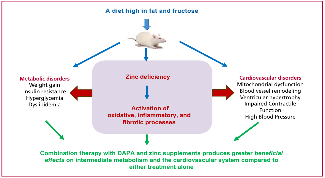
Therefore, we hypothesize that feeding a high-fat, high-fructose diet to male rats from weaning to adulthood promotes the development of MS and CVD by causing weight gain, dyslipidemia, hyperglycemia, oral glucose intolerance, hypertension, cardiac tissue remodeling, mitochondrial dysfunction in cardiomyocytes, and cardiac dysfunction. These alterations are mediated by increased oxidative, inflammatory, and fibrotic states.
Zinc supplementation helps reduce these alterations, given its antioxidant, anti-inflammatory, and insulinomimetic properties. Treatment with DAPA improves these abnormalities by inducing antihyperglycemic effects, lowering blood pressure, reducing oxidative and inflammatory processes, and improving mitochondrial function. Combining DAPA with zinc supplementation in rats with MS would produce greater beneficial effects on the intermediate metabolism and cardiovascular system compared with either treatment alone (Figure 1).
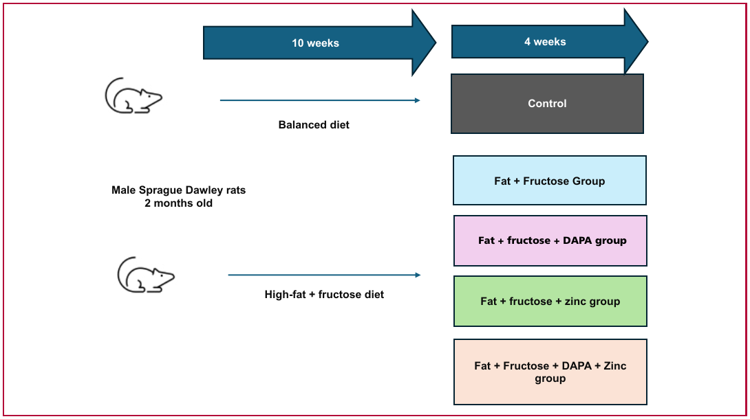
Based on this hypothesis, the objective of this study is to investigate the mechanisms by which the combined administration of zinc and DAPA, in a rat model of MS, could produce greater beneficial effects on the cardiovascular system and intermediate metabolism compared with either treatment alone. To achieve this objective, a basic research protocol is being conducted at the Department of Physiology of the School of Pharmacy and Biochemistry at Universidad de Buenos Aires (FFyB-UBA) and at the Institute of Chemistry and Drug Metabolism (IQUIMEFA)-UBA-CONICET, which has been approved by the CICUAL ethics committee of FFyB-UBA (REDEC-2023-26-E-UBA-DCT-FFyB), in accordance with the “Guide for the Care and Use of Laboratory Animals” (https://www.ncbi.nlm.nih.gov/books/NBK54050/) published by the National Academy of Sciences of the United States of America and updated by the American Physiological Society (APS) (https://www.physiology.org/career/policy-advocacy/animal-research?SSO=Y). For this study, male Sprague Dawley rats are fed either a balanced diet or a high fat (60% of total kcal) plus high fructose (10%) in ad libitum drinking water diet. After 10 weeks, the group fed the high-fat + fructose diet is administered orally for 4 weeks one of the following: a zinc supplement at 10 mg/kg/day; DAPA at 1 mg/kg/day; DAPA at 1 mg/kg/day plus a zinc supplement; or water (Figure 2). At the end of the experimental period, the effects on carbohydrate metabolism are assessed using an oral glucose tolerance test, and serum lipid profiles are analyzed. To evaluate the effects on the cardiovascular system, blood pressure measurements, echocardiographic studies, and morphological studies of the heart and its vessels are performed; the morphology and function of cardiac mitochondria are assessed. In addition, markers of fibrosis (transforming growth factor beta, TGF-β), inflammation (tumor necrosis factor alpha, TNF-α, and interleukin 6, IL-6), and oxidative stress (lipid peroxidation and the activity of the antioxidant enzymes superoxide dismutase, catalase, and glutathione peroxidase) in cardiac tissue are determined. To carry out this project, a research grant from the Argentine Society of Cardiology has been awarded in 2024 .
Discussion and Conclusion
The results of this research may represent a significant advance in addressing diseases of complex and multifactorial origin, such as MS. To date, there have been few basic research studies examining the relationship between DAPA and zinc. Anton I demonstrated that zinc supplementation in a rat model of diabetes—induced by streptozotocin and a high-fat diet—potentiates the effects of DAPA on metabolic conditions by producing greater reductions in blood glucose levels, improving liver function and lipid metabolism, and reducing oxidative stress compared with either treatment alone. (24) Furthermore, in a rat model of streptozotocin-induced diabetes, the combination of zinc with canagliflozin, another SGLT2i, has been shown to improve control of diabetes-related weight loss and hepatic steatosis. (25) Additionally, new DAPA-zinc complexes have demonstrated protective effects against liver damage and oxidative injury in diabetic rats. (22) Zinc plays important roles in lipid metabolism and insulin sensitivity through mechanisms involving ZAG and insulin signaling pathways, which may help enhance the effects of DAPA in MS. (11,20)
However, further basic and clinical research is needed to confirm and advance the investigation of zinc-DAPA combination effects on the various cardiovascular abnormalities associated with MS. Therefore, this study will help generate new therapeutic strategies for managing MS and preventing its progression to serious complications.
Financiamiento:
Research grant from the Argentine Society of Cardiology, Argentina, 2024–2025. Grant from Universidad de Buenos Aires, Argentina, 2023–2026, Code: 20020220100246B.