
Kounis syndrome (KS) is defined as the occurrence of an acute coronary syndrome in the setting of a hypersensitivity reaction. This represents a diagnostic challenge that extends beyond the traditional understanding of coronary artery disease. It was previously described as “allergic angina” due to its pathophysiology based on the activation of mast cells located in the arterial tunica adventitia and atherosclerotic plaques. (1) Over time, the understanding of this condition has shifted from a purely vasospastic phenomenon to one involving plaque erosion and rupture mediated by inflammatory processes.
Three clinical variants are recognized: type 1 is characterized by vasospasm in angiographically normal coronary arteries; type 2 involves erosion or rupture of a preexisting plaque, and type 3 presents as stent thrombosis. Recognition of type 2 is crucial because it involves the coexistence of stable chronic coronary artery disease and a concomitant immunologic trigger for instability, requiring prompt clinical management. (2,3) This manuscript presents a case of type 2 KS.
We report a case of a 68-year-old female patient, with appropriate informed consent. She had a history of hypertension, non-insulin-dependent type 2 diabetes mellitus, stage 3a chronic kidney disease, moderate to severe aortic insufficiency, two-vessel coronary artery disease treated with percutaneous coronary intervention (PCI) with stent placement in the circumflex and posterior descending arteries, with no further revascularization options as of 2018, and right knee osteoarthritis. She was admitted to a tertiary care center on an outpatient basis on September 6, 2025 for elective right knee replacement surgery. The procedure was performed without complications.
On September 7, 2025, during the immediate postoperative period (approximately 5:30 a.m.), following the administration of dipyrone (metamizole) for pain control, the patient developed a 5-minute episode of sudden, oppressive chest pain without radiation, associated with dyspnea, requiring oxygen support therapy via nasal cannula at 1 L/min. On room air (FiO2 0.21), arterial oxygen saturation was 88%. The electrocardiogram showed sinus rhythm at 85 bpm, regular R-R intervals, and Q waves in the inferobasal wall, without ST-segment elevation. Laboratory tests were performed, and cardiac biomarkers showed elevated troponin T on the second measurement (Table 1). A chest CT angiography using a pulmonary thromboembolism protocol was performed, and the result was negative. Transthoracic echocardiography showed a non-dilated left ventricle with concentric hypertrophy, preserved left ventricular systolic function, no segmental wall motion abnormalities, and an ejection fraction of 61%, as well as severe aortic regurgitation due to retraction and partial prolapse of the right coronary cusp, with no evidence of pulmonary hypertension. Transesophageal echocardiography confirmed these findings.
Table 1
Basal laboratory test results and following the onset of chest pain.
| Parameter | September 6, 2025 | September 7, 2025 |
|---|---|---|
| White blood cells | 17.66 x 10³/µL | 16.45 x 10³/µL |
| Neutrophils | 16.54 x 10³/µL | 14.71 x 10³/µL |
| Lymphocytes | 0.65 x 10³/µL | 0.69 × 10³/µL |
| Monocytes | 0.34 × 10³/µL | 0.95 x 10³/µL |
| Eosinophils | 0 x 10³/µL | 0 x 10³/µL |
| Basophils | 0.02 x 10³/µL | 0 x 10³/µL |
| MCV | 88.2 fL | 88.1 fL |
| Platelets | 275 x 10³/µL | 255 x 10³/µL |
| Hematocrit | 44.6% | 36.5% |
| Hemoglobin | 14.6 g/dL | 12 g/dL |
| Triglycerides | 98.6 mg/dL | |
| VLDL | 19.7 mg/dL | |
| HDL | 29.9 mg/dL | |
| LDL | 28.8 mg/dL | |
| Total cholesterol | 71.9 mg/dL | |
| HbA1C | 6.2% | |
| Troponin T* | 14.3 ng/mL | 77.7 ng/mL |
| Blood urea nitrogen | 31.6 mg/dL | |
| Creatinine | 1.76 mg/dL |
*Troponin delta values showed a significant increase, supporting the diagnosis of ongoing NSTEMI. MCV: mean corpuscular volume
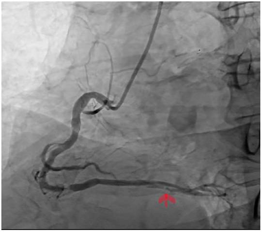
Based on these findings, a non-ST-segment elevation acute myocardial infarction was diagnosed, with a GRACE score of 128 points and suspected type 2 KS. Coronary angiography performed on September 19, 2025 revealed a severe lesion in the distal segment of the right coronary artery, with a previously implanted stent in a circumflex artery that was patent, without restenosis and with adequate flow; stent implantation was performed in the right coronary artery (Figure 1).
Following coronary angiography, the patient was transferred to the intensive care unit, with a favorable clinical course. Dual antiplatelet therapy and highintensity statins with ezetimibe were initiated. No evidence of impaired wound healing, infection, or discharge was observed; therefore, the orthopedic team opted for outpatient management.
This case highlights that a single dose of metamizole may destabilize a previously stable coronary plaque. In type 2 KS, the hypersensitivity reaction triggers massive mast cell degranulation. (4) This leads to the release of preformed mediators such as leukotrienes and histamine, which induce vasospasm, as well as proteases such as tryptase and chymase. These, in turn, activate matrix metalloproteinases that degrade collagen in the fibrous cap of the atherosclerotic plaque, facilitating erosion or acute rupture. In this patient, the presence of a severe distal lesion in the right coronary artery suggests inflammatory stress secondary to drug exposure, which acted as a trigger in a previously vulnerable coronary anatomy.
Metamizole (dipyrone) is a widely used analgesic, but its immunological safety profile requires close monitoring. (4) Agranulocytosis is the most widely recognized risk. However, this syndrome and immediate hypersensitivity reactions may frequently occur without evident cutaneous manifestations, as in this case. (5) A history of nonsteroidal anti-inflammatory drug (NSAID) intolerance is noteworthy, as it may indicate a pre-existing susceptibility to shift metabolism toward the leukotriene pathway.
From a therapeutic standpoint, KS remains a dilemma. Beta-blockers are contraindicated in the acute phase due to the risk of exacerbating coronary vasospasm. Adrenaline should be used with caution and reserved for anaphylactic shock, as it increases oxygen demand and may worsen ischemia. Therefore, a multidisciplinary approach involving cardiology is essential to guide revascularization along with corticosteroids and antihistamines. (6) This case underscores that, in the setting of an allergic reaction, the heart is also a critical target organ.
Conflicts of interest
None declared.
(See conflicts of interest forms on the website).
Ethical considerations
The authors declare that all procedures complied with institutional ethical standards and that patient confidentiality was ensured.