

Pulmonary sequestration is a rare congenital malformation, characterized by nonfunctional lung parenchyma, without communication with the tracheobronchial tree and with aberrant systemic arterial perfusion. Although the identification of an anomalous feeding artery usually guides the diagnosis, complex vascular variants can pose a diagnostic and therapeutic challenge. In this context, imaging techniques play a fundamental role in defining the vascular anatomy and planning treatment.
We present the case of a full-term newborn with a prenatal diagnosis of suspected left pulmonary sequestration, referred to a tertiary care center for evaluation and potential surgical resolution.
Postnatal thoracic ultrasound revealed an image consistent with pulmonary consolidation containing vascular structures. Subsequently, a chest computed tomography scan showed localized opacity in the left lower lobe.
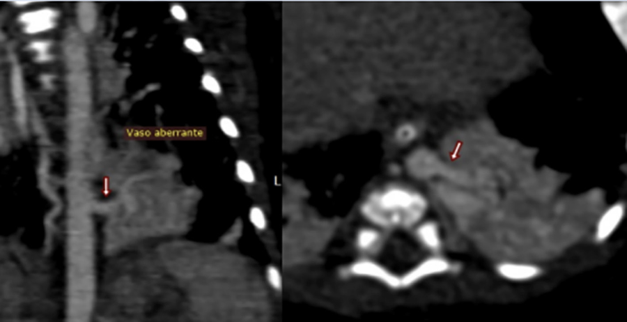
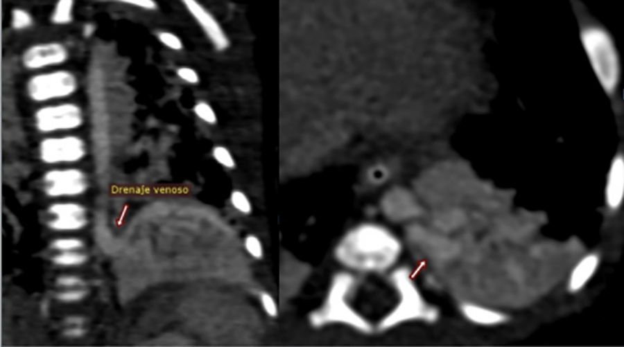
Thoracic angiotomography identified an aberrant arterial vessel originating from the descending aorta that supplied the lesion (Figure 1). In addition, it revealed an anomalous large-caliber venous drainage into the systemic venous system via a left paravertebral collector vein that drained into the innominate vein (Figure 2), a finding that is uncommon for this type of malformation.
To accurately characterize the vascular anatomy, an angiographic study was performed, which confirmed the presence of two systemic arteries originating from the descending thoracic aorta and supplying the left lower lobe (Figure 3). Furthermore, dual venous drainage was observed: one to the left atrium via the left inferior pulmonary vein (Figure 4) and the other to the innominate vein through the paravertebral collector (Figure 5), constituting a mixed venous return.
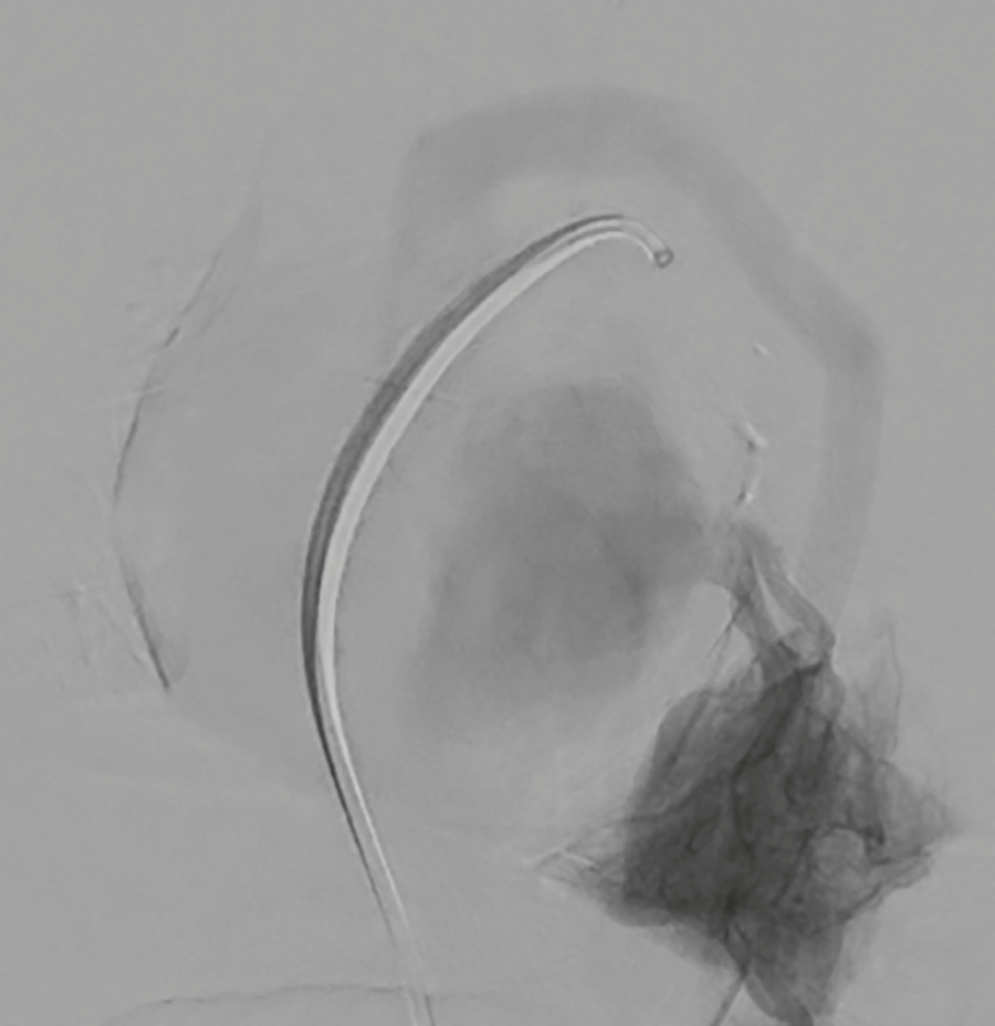
Fig. 4
Angiography. Venous phase. Venous drainage to the left atrium via the left inferior pulmonary vein
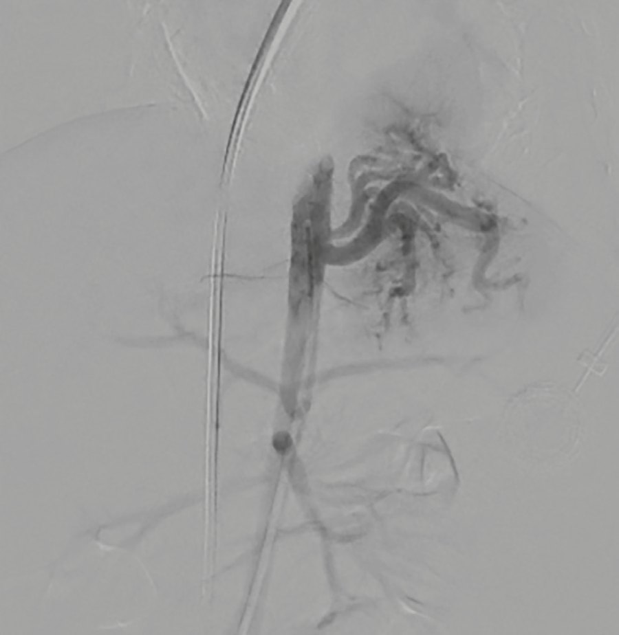
Fig. 5
Angiography. Venous phase. Venous drainage to the innominate vein through the paravertebral collector

A left lower lobectomy was performed, including dissection and control of the aberrant vessels, with a favorable postoperative course.
Pulmonary sequestration accounts for between 1% and 6% of congenital pulmonary malformations, with the left lower lobe being the most common site. (1) It is traditionally classified as intralobar or extralobar sequestration. The former shares the visceral pleura with the adjacent lung and usually drains into the pulmonary veins, whereas the extralobar type has its own pleural covering and typically drains into the systemic venous circulation. In addition, the latter may be associated with other congenital anomalies, such as diaphragmatic hernia, cardiovascular malformations, and gastrointestinal abnormalities. (2,3)
Systemic arterial supply is the defining feature of this condition. In most cases, the vascular supply originates from a single branch arising from the descending thoracic aorta; however, less common variants with multiple afferent vessels or originating from the abdominal aorta have been described. In our patient, angiography revealed the presence of two systemic arteries arising from the descending thoracic aorta, associated with a mixed venous drainage pattern. This combination constitutes a rare anatomical variant, which complicates the traditional classification of the lesion and underscores the need for a thorough anatomical evaluation before determining the therapeutic approach.
Currently, the diagnosis is often suspected through prenatal ultrasound, which achieves detection rates exceeding 80% for congenital pulmonary malformations. (3) In the postnatal period, chest X-rays and ultrasound may suggest the diagnosis, although definitive characterization of the vascularization requires advanced studies such as computed tomography angiography or magnetic resonance imaging. When di- agnostic uncertainty persists or a precise anatomical definition is required for treatment planning, angiography remains an invaluable tool.
In this case, angiotomography made it possible to identify the aberrant arterial supply and detect unexpected systemic venous drainage, while angiography provided a detailed characterization of the afferent and efferent vessels. This information proved essential for surgical planning, as it allowed for the identification of complex vascular anatomy and minimized the risk of intraoperative complications. The importance of this preoperative assessment lies in historical reports of morbidity and mortality associated with inadvertent injury to aberrant vessels during surgical resection. Although the surgery has a low overall complication rate, close to 5%, intraoperative bleeding remains one of the most significant adverse events, followed by hemothorax and bronchopleural fistula. (3)
Surgical resection remains the strategy of choice in symptomatic patients and in most intralobar sequesters, due to the risk of recurrent infections. Alternatively, embolization of aberrant systemic vessels has emerged as a valid therapeutic option in selected cases, especially when significant shunts or heart failure are present. (1,4) In our patient, the complexity of the vascular anatomy and the presence of mixed venous drainage made surgical resection the most appropriate option, with a favorable postoperative course.
This case highlights the value of imaging techniques in the evaluation of pulmonary sequestration, especially when unusual anatomical variants are pre- sent. The thorough characterization of arterial perfusion and venous drainage allowed us to identify unexpected vascular anatomy and appropriately guide the surgical strategy, contributing to a favorable outcome.