

We present the case of a patient with stage IV lung adenocarcinoma whose clinical presentation was cardiac tamponade secondary to pericardial effusion. Although pericardial effusion is a relatively common complication in patients with cancer, only about 30% of cases are attributable to malignant pericardial infiltration, (1) making its accurate characterization essential due to its diagnostic, prognostic, and therapeutic implications. (2) The case is also notable for the complete resolution of the echocardiographic findings following the initiation of targeted systemic treatment, a phenomenon rarely described in the literature and which highlights the benefit of current oncological therapies.
The patient is a 46-year-old man, an active smoker (30 pack-years), with no other relevant personal medical history, who presented to the emergency department with progressive dyspnea, orthopnea, and pleuritic chest pain that had been present for one month, with clinical worsening and the onset of edema in the left lower extremity within the past 24 hours. Upon arrival, he presented with hypotension (94/65 mmHg), sinus tachycardia (120 bpm), tachypnea (30 bpm), and an oxygen saturation of 92% while breathing room air.
On suspicion of deep vein thrombosis with secondary pulmonary thromboembolism, an urgent thoracic computed tomography (CT) angiography was performed (Figure 1A), which confirmed bilateral pulmonary thromboembolism with associated pulmonary infarction, as well as an unidentified mass in the right upper lobe measuring 25 mm, and another in the anterior mediastinum (60 mm), which was infiltrating the pericardium. The latter showed diffuse nodular thickening suggestive of pericardial carcinomatosis with severe pericardial effusion. A Cardiology evaluation was requested, and a transthoracic echocardiogram confirmed severe pericardial effusion and findings consistent with cardiac tamponade, including respiratory variation in transvalvular flows , a dilated and non-collapsible inferior vena cava, and evident diastolic collapse of the right ventricular chambers.
Fig. 1
Thoracic CT angiography. A) Pericardium showing irregular thickening and associated severe pericardial effusion. B) Complete resolution of the pericardial effusion and marked reduction in pericardial infiltration at 3 months.
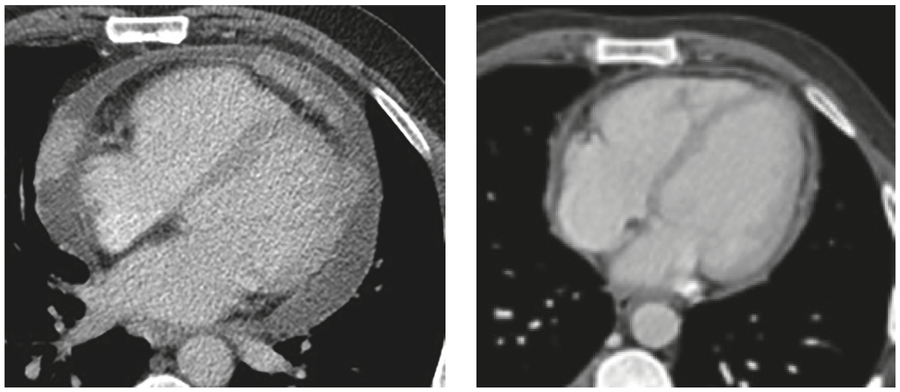
Given these findings, the patient was admitted to the Coronary Care Unit, where an emergency pericardiocentesis was performed. A total of 500 mL of serosanguineous fluid was obtained. Subsequently, anticoagulation with low-molecular-weight heparin was initiated to treat pulmonary thromboembolism. Although he initially showed clear clinical and hemodynamic improvement, he continued to present with high-volume pericardial drainage, persistent severe pericardial effusion, and very marked nodular pericardial thickening (Figure 2A). Although the cytological examination of the pericardial fluid was negative for malignant cells, the findings on CT angiography were highly suggestive of malignant infiltration: (3) loss of the fatty plane separating the pericardium from the adjacent mediastinal mass, and nodular pericardial thickening. It was ultimately decided to perform a percutaneous balloon pericardiotomy, which stabilized the patient’s condition and allowed for hospital discharge a few days later.
Fig. 2
Transthoracic echocardiogram in the parasternal long-axis view. A) Irregular pericardial thickening with associated severe pericardial effusion (with persistence of variationtransvalvular respiratory, but without diastolic involvement of the right chambers following pericardiocentesis). B) Resolution of pericardial thickening and effusion at 3 months.
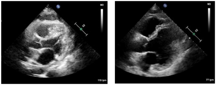
The pathological examination confirmed the diagnosis of stage IV A pulmonary adenocarcinoma. After ruling out abnormalities in molecular biomarkers (EGFR, ALK, ROS1, BRAF) and confirming the expression of programmed cell death ligand 1 (PD-L1) —which was not amenable to targeted therapy—firstline palliative treatment with pembrolizumab, pemetrexed, and carboplatin was initiated.
At the three-month follow-up, the patient showed a favorable clinical course, with a partial response characterized by a reduction in the mediastinal mass to 32 mm, the lobar mass to 18 mm, and a significant reduction in pericardial infiltration, with minimal pericardial effusion on the follow-up CT scan (Figure 1B) and complete resolution of the findings suggestive of pericardial infiltration visible on echocardiography (Figure 2B).
The clinical presentation of this patient, who presented with a pericardial effusion and cardiac tamponade, leading to a diagnosis of advanced-stage lung adenocarcinoma, is not an exceptional situation in clinical practice. An underlying neoplasm has been reported in 12–23% of patients who present with a pericardial effusion as their initial manifestation. (4) Among malignant etiologies, lung cancer is one of the most common causes, along with breast cancer, melanomas, leukemias, and lymphomas. (1,4)
Although a definitive diagnosis of malignant pericardial effusion requires confirmation of tumor cells, the diagnostic sensitivity of cytology is estimated at 72%, (5) and pericardial biopsy is rarely performed. In cases such as this one, where false negatives in cytology are suspected, it is vital to integrate the findings from clinical and various imaging tests to arrive at the most likely diagnosis and adapt the therapeutic management accordingly.
Pericardial effusion of neoplastic origin is associated with a high recurrence rate and an increased risk of cardiac tamponade. (4) For this reason, in addition to pericardiocentesis, complementary strategies may be necessary, such as percutaneous balloon pericardiotomy, creation of a pericardial window, or intrapericardial administration of cytostatic agents, especially in patients with early recurrence. (1) However, treatment of the primary tumor remains the cornerstone for controlling the disease and its cardiovascular manifestations. (1)
The incorporation of immunotherapy and current systemic treatments has transformed the prognosis for many patients with advanced lung cancer, improving both survival and quality of life. In our patient, the partial radiological response was accompanied by the complete resolution of the pericardial effusion and of the echocardiographic findings suggestive of pericardial infiltration—a finding not previously described in the literature to the best of our knowledge—which illustrates the efficacy of systemic treatment and the value of echocardiography as a noninvasive tool for monitoring pericardial involvement.