

Thoracic operculum syndrome (TOS) is defined by extrinsic compression of the brachial plexus and/or subclavian vessels due to the presence of bony abnormalities or hypertrophy of the scalene and/or subclavian muscles. Involvement of the subclavian artery is rare but serious, due to the risk of thromboembolic complications such as acute arterial ischemia of the upper limb or posterior stroke caused by retrograde embolism. (1)
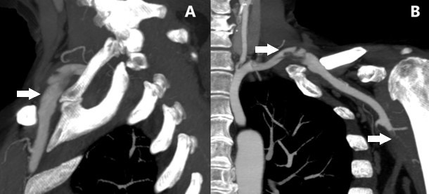
We present the case of a 66-year-old woman with a history of poliomyelitis and residual paresis of the left lower limb who used an orthosis and a forearm crutch to walk. She presented to the emergency department with pain in her left shoulder radiating to her forearm, accompanied by paresthesias that had been present for 24 hours. On physical examination, she presented with coldness and cyanosis, absence of radial and ulnar pulses in the left upper limb, with preserved sensation and motor function. The ECG showed a sinus rhythm; the chest X-ray revealed bilateral cervical ribs (Figure 1A); and the echocardiogram showed mild dilatation of the left atrium. Vascular Doppler ultrasound revealed a partially thrombosed aneurysm of the left subclavian artery and thrombosis of the humeral artery, with monophasic flow in the radial and ulnar arteries. Anticoagulation was initiated with intravenous unfractionated sodium heparin via an infusion pump. Computed tomography angiography (CTA) demonstrated thrombosis of the left humeral artery; bilateral supernumerary ribs at the C7 vertebral segment, with a fibrocartilaginous junction to the first left sternal rib, which exerted extrinsic compression on the subclavian artery aneurysm, resulting in right-sided vascular compromise. (Figure 1B and Figure 2)
Fig. 1
A) Chest X-ray: bilateral cervical ribs. B) CT angiography of the thoracic aorta and upper extremities: extrinsic compression of the subclavian artery by a cervical rib (arterial TOS)
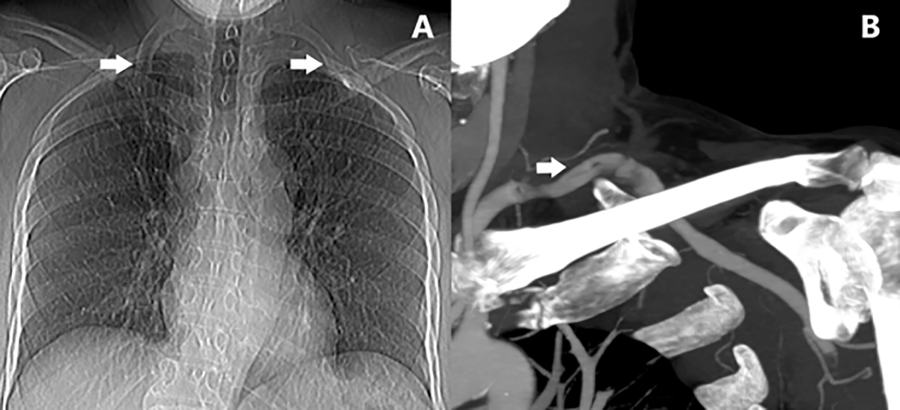
Fig. 2
A) CT angiography of the thoracic aorta and upper extremities: compression of the subclavian artery due to pseudoarthrosis of the left cervical rib with the first sternal rib. B) CT angiography of the thoracic aorta and upper extremities: partially thrombosed subclavian artery due to TOS, associated to humeral artery thrombosis due to distal embolism.
An emergency thromboembolectomy was performed using a Fogarty catheter on the subclavian, axillary, and humeral arteries via an approach in the middle third of the arm. Postoperatively, the radial pulse was restored, and anticoagulation with heparin was resumed.
During hospitalization, decompressive surgery was performed on the TO via a supraclavicular and infraclavicular approach, including resection of the cervical rib and first sternal rib, anterior and middle scalene resection, and resection of the subclavian aneurysm with arterial reconstruction using a prosthetic subclavian-axillary bypass (6-mm ringed polytetrafluoroethylene graft). Anticoagulation with acenocoumarol was prescribed for 3 months. At the one-year followup, the patient remained asymptomatic with a radial pulse, and CT angiography showed a patent bypass.
Acute arterial ischemia in the upper extremities is less common than in the lower extremities due to the extensive collateral circulation around the shoulder and elbow.
Jo et al., in a review of 366 cases, reported a thrombotic cause in 61.7% and an embolic cause in the remainder. The most common causes included thrombosis of a vascular access site and cardiac embolism due to atrial fibrillation. The humeral artery is the most frequently affected, either alone or in combination with other arteries, in 89% of cases. (2)
Arterial TOS is characterized by compression of the subclavian artery, most commonly by a cervical rib or other bony anomalies such as a hypoplastic first rib, clavicle fracture, or hypertrophic calluses from fractures. Lesions in the subclavian artery result from chronic compression that leads to changes in the arterial wall, including intimal injury, ulcers with or with- out post-stenotic dilation, aneurysms, and occlusive thrombosis. It is the least common form of thoracic operculum, accounting for only 3–4 ,4% of cases, and is most commonly described in young adult women. (1,3,4)
The clinical presentation of arterial TOS is diverse, secondary to the broad spectrum of associated bone and arterial pathology. Cases have been described of intermittent claudication, digital ischemic necrosis, acute upper extremity arterial ischemia, posterior stroke, as well as overlap with symptoms of neurogenic TOS and asymptomatic cases identified on imaging studies. (1,3,5,6)
Acute upper extremity arterial ischemia has been reported in 15–30% of cases of arterial TOS, commonly due to embolization from an aneurysmal and thrombosed subclavian artery. (1,3,5)
Diagnostic methods include chest X-ray, which identifies bony abnormalities, and Doppler ultrasound to visualize arterial aneurysms or stenoses, as well as thrombosis and distal embolism. CT angiography provides anatomical details of the bony abnormality, subclavian artery involvement, and distal vascular beds for revascularization. Angiography is recommended when the diagnosis is unclear or to confirm subclavian artery compression in the early stages. (3,6)
The initial management of acute limb-threatening ischemia includes surgical thromboembolectomy with a Fogarty catheter or percutaneous thrombolysisthrombectomy, followed by systemic anticoagulation with heparin. (1,3,4)
Pitcher et al., in a 30-year retrospective analysis, reported that 62% of patients with acute arterial ischemia due to arterial TOS experienced a recurrent thromboembolic event, suggesting that decompressive surgery of the operculum during hospitalization may help prevent it. (5)
Decompressive treatment for arterial TOS includes anterior and mid-scalenectomy with resection of the cervical rib and/or first rib or other bony abnormality in all cases, while vascular reconstruction is performed selectively based on the severity of subclavian artery disease. (1,3,5) The reported reconstruction rate ranges from 38% to 70%, with procedures including saphenous vein bypass, prosthetic bypass, or cryopreserved vessel bypass; resection with endto-end anastomosis; or surgical angioplasty with a venous patch. (1,5) In subclavian arteries with mild post-stenotic dilations without intimal disruption, vascular reconstruction is not indicated, and followup with Doppler ultrasound is recommended. (1,5)
The supraclavicular approach is the most commonly used for decompression, and when arterial reconstruction is required, a combined supraclavicular and infraclavicular (paraclavicular) approach is preferred for better vascular exposure. (1,3,5)
In cases of acute arterial ischemia due to TOS, lifelong antiplatelet therapy and anticoagulation for 3 to 6 months are recommended. (5)
Stent angioplasty is not recommended as a primary treatment for arterial SOT, as it does not address the cause of the arterial pathology, and bone compression can lead to stent fracture or thrombosis. (3)
The vascular manifestations of arterial TOS carry the greatest potential for disability, as 30% of patients present with persistent symptoms such as chronic pain and related to the degree of thromboembolism in the distal arteries prior to decompressive treatment. (1-3,5)
In summary, TOS is a rare cause of arterial compromise in the upper extremity. In cases of acute arterial ischemia, thromboembolectomy and systemic anticoagulation are initially recommended, followed in all cases—and during the same hospital stay—by early decompressive surgery of the operculum to prevent recurrence of a thromboembolic event. Vascular reconstruction is performed selectively based on the extent of subclavian artery involvement.