

INTRODUCTION
Heart failure with reduced left ventricular ejection fraction (LVEF) represents one of the pathologies with the highest mortality. Among its main etiologies is dilated cardiomyopathy (DCM). The predictors of DCM development and prognosis are subject to ongoing research. Despite advances in understanding the underlying pathophysiological mechanisms, the reasons why certain conditions predispose to the progression of dilated cardiomyopathy are still not fully understood. (1)
Genotypic (familial) alterations that are responsible for a small group of cases with the ventricular dilatation phenotype have been identified.
A notable aspect is that the same noxa can generate variable effects among different patients. This suggests the possible existence of a genetic predisposition that would influence a differentiated clinical evolution. In this context, single nucleotide polymorphisms (SNPs) could play a key role in the variability of patients’ response to the same noxa.
To investigate the existence of genetic variants associated with the predisposition to DCM, we used Chagas disease as model. (2) Chagas disease is responsible for DCM with the highest morbidity and mortality. It presents two clinical phases: an acute phase, which is usually asymptomatic in 95% of patients, and a chronic phase, with a long latency period of 10-30 years. In this group, between 25% and 30% of individuals develop signs or symptoms of cardiac involvement (Chagas cardiomyopathy). (3)
Chronic Chagas cardiomyopathy (CCM) has been considered the most frequent cause of non-ischemic cardiomyopathy in Argentina. However, there is no consensus on the risk indicators for the development of this condition and other cardiovascular events, regardless of the effect of traditional risk factors. (4)
OBJECTIVE
The aim of our study was to determine the genetic variants related to the presence of DCM by performing an association analysis with SNPs.
METHODS
Prospective single-center study that included patients from June 2016 to January 2020.
Patients from 21 to 80 years of age, seropositive for Chagas disease with DCM, from the provinces of northwestern Argentina and southern Bolivia, and other serologically positive patients of similar age, from the same geographic areas without DCM, were included as a control group.
All patients should have a known history of infection of more than 20 years certified by the treating physician, serological analysis, and abandonment of the endemic area for more than 20 years as well as a LVEF measured within 12 months prior to inclusion, ≤ 35% or ≥ 50%.
Patients who refused to sign the informed consent form, those with known serious pathologies (excluding cardiovascular) that generate a life expectancy of less than one year, and those who were participating in research protocols in the 30 days prior to sample collection were excluded. Those in whom personal or telephone contact could not be assured, with alcohol or drug abuse in the last 6 months, with clinical and laboratory evidence of liver failure (transaminase value x3 and total bilirubin >2 mg/dL), with LVEF between 36% and 49% (the objective was to separate the groups avoiding patient crossover), and those receiving drugs with a known action on cardiovascular parameters (immunosuppressants, nitrates, estrogens) were also rejected. Patients with acute or chronic coronary artery disease, with an indication for revascularization or with coronary angioplasty or myocardial revascularization surgery in the previous 6 months, patients with renal failure (serum creatinine >2.5 g/dL), with severe obstructive pulmonary disease, with DCM of other etiologies (e.g. rheumatoid arthritis, diabetes, hypertension), with significant valvular diseases (except those secondary to mitral and/or tricuspid valve annulus dilatation), with pacemakers with stimulation greater than 50%, or with autoimmune pathologies (lupus, scleroderma, hepatitis C, etc.) were also excluded.
Patients who met the inclusion criteria and did not present exclusion criteria were divided into 2 groups:
Group 1: patients with LVEF≤ 35%.
Group 2: individuals with LVEF≥50%.
After signing the informed consent, a complete clinical history and physical examination were performed, according to their underlying pathology and the corresponding diagnostic and prognostic procedures.
An echocardiogram to confirm the LVEF value and a 12-lead ECG were performed. According to the ECG results, patients were classified into two groups: with intraventricular conduction disorders (IVCD), when they had one or more of the following criteria: right bundle branch block, left bundle branch block, left anterior hemiblock and left posterior hemiblock; or without IVCD.
Patients were coded with an alphanumeric system to ensure the identity of the groups.
A 10 mL blood sample was taken by trained personnel for genomic determination. The sample was placed in tubes with the presence of ethylenediaminetetraacetic acid (EDTA) according to ISO standards to prevent coagulation and stored at -20ºC until processing. This sample was processed to obtain the corresponding deoxyribonucleic acid (DNA) in a specialized center in our country, leaving safety samples.
The DNA was sent via a courier authorized by international legislation for this purpose to the international laboratory Xenética Cardiovascular, Instituto de Investigación Sanitaria de Santiago, laboratory nº1, Complexo Hospitalario Universitario de Santiago de Compostela, where genotyping and analysis were performed (Table 1). The laboratory remained blind to which group the patients belonged. This was only known by the statistical team at the time of the analysis.
An SNPs panel of genes was prepared related to the following:
Ventricular contraction and relaxation process (TTN, BAG3, MTSS1). (5, 6, 7)
Myocardial metabolism (PPARGC1A, SIRT1, AKT1 mTOR, AMPK, PRKAA2).
Beta 1 adrenergic receptor (ADRB1). (8, 9, 10, 11)
Cholinergic receptor muscarinic 2 (CHRM2).
Angiotensin II type 1 receptor (AGTR1B).
Atrial natriuretic peptide (NPPA).
Cell cycle regulators (CDKN1A, RYR2)
Sarcomeric structure (ATP2A2, DSP, JUP)
Cellular energy (PRKAB2, PRKAB1)
Mitochondrial activity (SOD 2)
Apoptosis (AKT1)
Cellular hypoxia regulator (HIF1A)
Respiratory chain (LDHA, lactate dehydrogenase (LDH))
Nitric oxide (NOS1, NOS2P3)
Growth and inflammation factors (SH2B3)
The international database TheGenomeAggregationDatabase (GnomAD) was used, choosing SNPs with a frequency of 10% to 40% in the population incorporated into this database.
Finally, 68 SNPs, detailed in Table S1, were selected for analysis.
The technology used was iPLEX Gold. The script was created in SNPassoc to evaluate whether the entire population met the H-W equilibrium condition.
The association between these two phenotypes was evaluated as binomial variables coded as YES=1 and NO=0, with the SNPs coded as an ordinal variable according to the following convention: 0 for homozygotes of the wild-type allele, 2 for homozygotes of the alternative allele and 1 for heterozygotes.
To evaluate the relationship between the different SNPs under study and the presence of reduced LVEF, various artificial intelligence models for supervised learning were used. The performance of 1) Logistic Regression, 2) Support Vector Machines, 3) Artificial Neural Networks, 4) Naive Bayes, 5) Decision Trees and 6) Random Forest was evaluated, with the collaboration of the genomIT Artificial Intelligence team.
The performance evaluation of the different models for the prediction of rEF was carried out using the cross-validation technique, using F1 score as a measure of precision to select the best model.
To evaluate the relationship between each SNP and the presence of IVCD, a univariate logistic regression model with the presence of reduced LVEF and IVCD as outcomes and each SNP as a predictor was evaluated. SNPs with an associated p value<0.05 were selected for evaluation of their effect using a multivariate logistic regression model, selecting those SNPs with a p value<0.05 using the backward selection strategy with stepwise regression method. The magnitude of the association between each SNP and the outcome under study was expressed as the Odds Ratio and its respective 95% confidence interval (95% CI). The predictive performance of the model was evaluated using the area under the curve (AUC). The same procedure was applied using logistic regression of the SNPs with reduced LVEF.
The analyses were performed with the R ® software (version 4.1.1, R Development Core Team/R Foundation for Statistical Computing, Vienna, Austria).
A Manhattan plot was also generated with different inheritance models for all SNPs, analyzing the nominal level of significance and the Bonferroni level.
Ethical considerations
The study was developed in accordance with the ethical principles of the Declaration of Helsinki and approved by the Teaching and Research Committee of the institution.
RESULTS
One hundred and eighty-two patients with positive serology for Chagas disease from southern Bolivia and northwestern Argentina were studied.
The median age of the population was 62 years, 39.6% were men. Fifty-seven of them (31%) had rEF and 95 (53%) had some IVCD (Table 1).
Table 1
Baseline characteristics
| Preserved LVEF | Reduced LVEF | |
|---|---|---|
| N | 125 | 57 |
| Age (years) | 59.44 (11.25) | 64.68 (10.09) |
| Male gender | 39 (31.2) | 33 (57.9) |
| HTN | 38 (30.4) | 13 (22.8) |
| DM | ||
| No | 116 (92.8) | 51 (89.5) |
| Type I | 3 (2.4) | 0 (0.0) |
| Type II | 6 (4.8) | 6 (10.5) |
| HF | 4 (3.2) | 34 (59.6) |
| Arrhythmias | 21 (16.9) | 28 (50.0) |
DM: diabetes mellitus; HF: heart failure; HTN: hypertension; LVEF: left ventricular ejection fraction
Quantitative variables are presented as mean and standard deviation, qualitative variables as frequency and percentage.
Among the different supervised classification models evaluated, the model that uses 7 K folds and a logistic regression classifier obtained a score of F1=0.85, being the best model obtained; therefore its results are reported.
The univariate analysis of the relationship between each SNP (on the horizontal axis) and the presence of a reduced LVEF and IVCD respectively performed by logistic regression is presented in Figures 1 A and 1 B.
Figures 1
A and B: The vertical axis expresses the p values for each SNP, the red line marks a p = 0.05 significance level, and the red dots correspond to the SNPs that presented a p value < 0.05.

IVCD: intraventricular conduction disorder; SNP: single nucleotide polymorphism
A multivariate analysis that revealed the association of reduced LVEF phenotype and IVCD was performed. (Figures 2 A and B, Tables 2 and 3).
It was observed that the variants rs2076300, rs61772962 and rs7071853 were independent predictors of reduced LVEF; whereas, for the presence of blocks, the only predictor was the SNP rs72840788, as is seen in the univariate analysis.
Figures 2
A and B. Odds ratio and 95% CI for SNPs predicting A) reduced LVEF and B) IVCD phenotype.

95% CI: 95% confidence interval; IVCD: intraventricular conduction disorder; LVEF: left ventricular ejection fraction; SNP: single nucleotide polymorphism
Table 2
Multivariate analysis. Odds ratio and 95% CI and p-value for each of the three SNPs independently associated with reduced LVEF.
| Reduced LVEF | |||
|---|---|---|---|
| Predictor | Odds ratio | 95% CI | p value |
| rs2076300 | 1.79 | 1.01 - 3.21 | 0.046 |
| rs61772962 | 1.77 | 1.00 - 3.14 | 0.049 |
| rs7071853 | 1.77 | 1.07 - 2.96 | 0.026 |
95% CI: 95% confidence interval; LVEF: left ventricular ejection fraction; SNP: single nucleotide polymorphism
Table 3
Odds Ratio, 95% CI and p value for the only SNP associated with the presence of intraventricular conduction disorders
| Intraventricular conduction disorders | |||
|---|---|---|---|
| Predictor | Odds ratio | 95% CI | p value |
| rs72840788 | 2.44 | 1.04 - 6.22 | 0.048 |
95% CI: 95% confidence interval; SNP: single nucleotide polymorphism
In each of the three SNPs predicting DCM, the proportion of homozygous wild-type, heterozygous and each homozygous variantof the alternative allele was analyzed, according to the presence or not of a reduced LVEF and IVCD. (Figures 3 A, B and C, Figure 4)
The predictive performance of the model for the presence of reduced LVEF is evidenced by the area under the ROC curve (0.662) and its corresponding 95% CI (0.580-0.745) (Figure 5)
Figure 3
Proportion of homozygous wild-type (0), heterozygous (1) and homozygous alternative allele (2) variants of SNP rs2076300 (3 A), rs61772962 (3 B) and rs7071853 (3 C), according to the presence of reduced or preserved LVEF
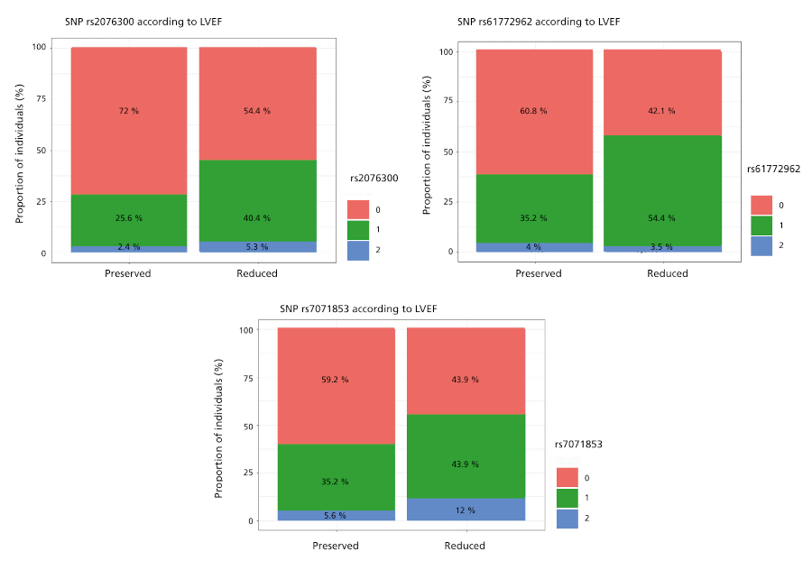
LVEF: left ventricular ejection fraction; SNP: single nucleotide polymorphism
0: homozygous for the wild-type allele 1: heterozygous 2: homozygous for the alternative allele.
Figure 4
Proportion of homozygous wild-type, heterozygous and homozygous variants of the alternative allele of SNPrs72840788, according to the presence of IVCD
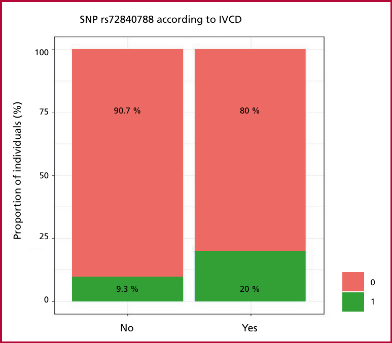
IVCD: intraventricular conduction disorder; SNP: single nucleotide polymorphism
0: homozygous for the wild-type allele 1: heterozygous
Figure 5
Predictive performance of the model for the presence of reduced LVEF c the area under the ROC curve (0.662) and its 95% CI (0.580-0.745)
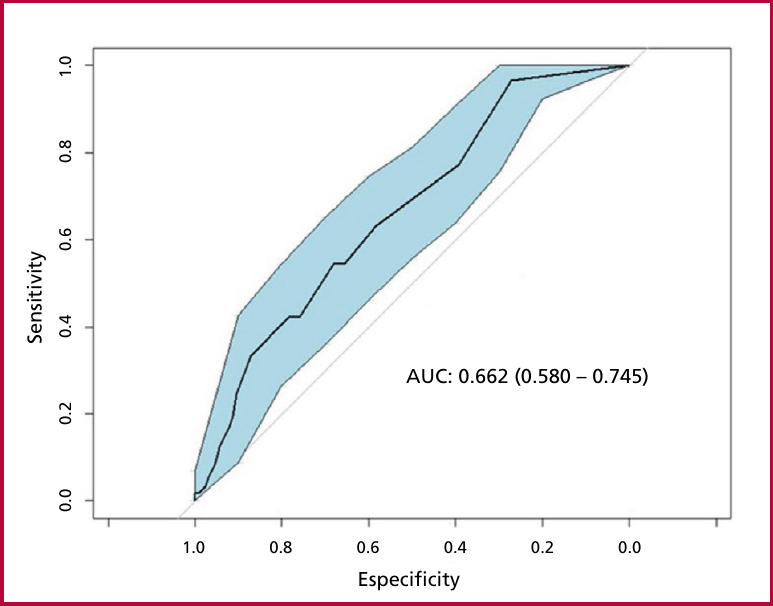
AUC: area under the curve; LVEF: left ventricular ejection fraction; 95% CI: 95% confidence interval; SNP: single nucleotide polymorphism
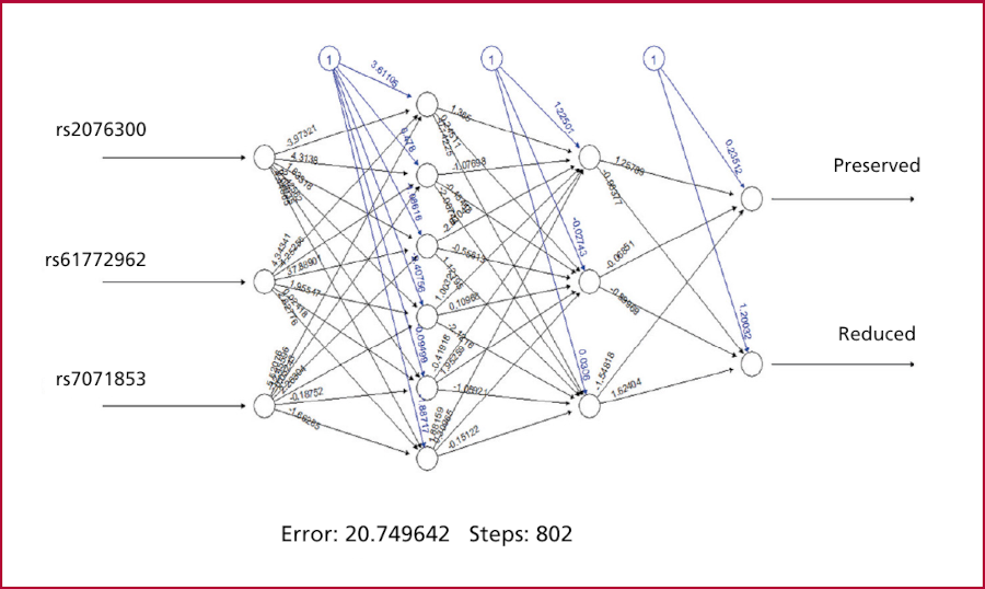
Regarding the performance of the neural network for the prediction of reduced LVEF with the three identified SNPs, the best model was that of a neural network with 2 hidden layers of 6 and 3 neurons respectively, with an error rate (proportion of prediction failures) of 0.33 and an accuracy rate (proportion of prediction successes) of 0.67, (Figure 6).
DISCUSSION
Heart failure (HF) is a disease of epidemic proportions, representing the final stage of various pathologies. Its prevalence increases two to three times when including patients with asymptomatic systolic dysfunction. (12, 13, 14)
Initially, 60-70% of patients with HF had a high mortality within five years after diagnosis, accompanied by a high rate of hospitalization for decompensated HF. However, with the advent of new drugs, a significant decrease in morbidity and mortality has been seen. (15, 16, 17, 18, 19, 20, 21)
Heart failure with reduced LVEF is a progressive disorder that usually begins after a noxa or damaging event that affects the heart muscle, resulting in the loss of functional myocytes or a decrease in the contractile capacity of the myocardium. In most cases, an asymptomatic phase initially occurs due to compensatory mechanisms that are activated and modulate LV function within physiological limits for a certain period of time. However, the sustained activation of the neurohormonal and cytokine systems causes a series of changes in the myocardium, leading to ventricular remodeling and the eventual appearance of symptomatic HF.
Neurohumoral systems include the sympathetic nervous system (SNS), through the activation of the Beta 1 receptor, and the renin-angiotensin system (RAS), mediated by the persistent action of angiotensin II (AT2) and aldosterone on the myocardium.
Parasympathetic inhibition also contributes to HF pathogenesis by reducing nitric oxide (NO) concentrations with the consequent increase in inflammation and ventricular remodeling. (22)
Most of the genes involved in the development of DCM and HF are involved in energy production and regulation processes, as well as calcium signaling and transcription regulation. The genes described are mainly those of sarcomere proteins (MYH7, TNNT2, TNNI3, TNNC, TPM1, MYBPC3, TTN, ACTC, MYL3, MYL2), Z-disc proteins (VCL, LDB3, TCAP, MYOZ2), cytoskeletal or cell membrane proteins (DES, DAG1, SGCA, LMNA, FLNC), desmosomal proteins (JUP, DSP, DSG2, DSC2, PKP2, RYR2, PLN), sodium channel (SCN5A), metabolic proteins (PRKAG2, GLA, LAMP2, GAA) and regulatory proteins (RMB20, BAG3, TGFB3) among others.
Through genome-wide association studies (GWAS), certain SNPs associated with the risk of developing HF have been described.
One of the most widely studied SNPs is the variant responsible for the change of arginine to glycine (Arg389Gly) in the Beta 1 receptor. In the general population, Arg389Gly polymorphism was not significantly associated with HF, but in the subgroup analysis according to ethnicity, the presence of Gly389 in Asian patients was shown to increase the risk of HF by 35% compared to Arg389 carriers (RR 1.35; 95% CI: 1.16-1.57; p < 0.001). In a more detailed subgroup analysis, Arg389 homozygotes were associated with an improvement in LVEF in East Asians (95% CI 1.85-3.40; p < 0.001) and mixed population (95% CI 0.72-2.91; p < 0.001); while among white patients, Arg389 homozygotes had an improvement in LV systolic diameter. The improvement was significantly greater than that of Gly389 (95% CI 0.04-0.36, p=0.001). (23-26)
In the case of renin genes, G/AI 9-83 polymorphisms were analyzed in patients with DCM. The heterozygous form was found in only 37.5% of control subjects. This study did not show involvement of renin A/G polymorphisms in HF pathogenesis. (27)
Patients with DCM may present hypertrophy and fibrosis. An association of the angiotensin II type 1 receptor and gene polymorphisms (AGTR1, A1166C) was observed in angiotensin converting enzyme (ACE)-mediated left ventricular hypertrophy (LVH) in endurance athletes, that would be a variant for future study in DCM. (28)
On the other hand, in several studies, two SNPs (rs763361 and rs727088) in the last exon of CD226 have been reported in the Titin gene (TTN), as associated with an increased risk of DCM. (29)
Another gene to consider is MMP2. This gene SNPs were analyzed to evaluate their association with the development of DCM and three of them were found to be related both to the risk of suffering from it as to its worse prognosis. (30)
Since there is no consensus on the variants that could predispose to the development of DCM in response to harmful factors affecting the myocardium, we decided to establish an analysis panel. This includes structures related both to neurohumoral systems as to aspects of energy metabolism that could be involved in the development of DCM. The international database TheGenomeAggregationDatabase (GnomAD) choosing SNPs with a frequency of 10% to 40% in the world population. The SNPs used are detailed in Table S1.
In our analysis we were able to observe that the variants rs61772962, rs7071853 and rs2076300 were independent predictors of reduced LVEF in our population of patients with chronic Chagas cardiomyopathy.
The SNP rs61772962 corresponds to an intronic variant of the PRKAA2 gene. This gene encodes the alpha-2 catalytic subunit of the 5'-AMP-activated protein kinase. This AMPK protein, is an important cellular energy-sensing enzyme. In response to cellular metabolic stress, AMPK is activated and thus phosphorylates and inactivates acetyl-CoAcarboxylase (ACC) and beta-hydroxy beta-methylglutaryl-CoAreductase (HMGCR) key enzymes involved in the regulation of de novo biosynthesis of fatty acids and cholesterol. (31) The AMPKα2 variant is required for energy metabolism in cardiomyocytes. (32)
The RNF207 and PRKAA2 genes, known for their involvement in cardiac action potentials, energy homeostasis and morphology, have been postulated as candidates for DCM models after study in dogs and follow-up in humans. (33)
Dominant mutations in the γ2 regulatory subunit of AMP-activated protein kinase (AMPK), encoded by the PRKAG2 gene, cause glycogen storage cardiomyopathy in mutant mice. (34)
The rs7071853 corresponds to the gene for the BAG3 regulatory protein (position chr10:119552094 (GRCh38.p14). On the other hand, rs72840788 was associated with the prediction of conduction disorders and corresponds to the intronic position of that protein. The BAG3 protein plays an important role in maintaining myocardial homeostasis and excitation-contraction coupling and is an adaptive mechanism to maintain cellular homeostasis under stress. It is most prominently expressed in the heart, skeletal muscle, and in many forms of cancer. In the heart, it acts as a chaperone for heat shock proteins to facilitate autophagy. Mutations in BAG3 have been associated with the development of a variety of phenotypes, including hypertrophic/restrictive and dilated cardiomyopathy. (35, 36, 37, 38, 39, 40)
The rs2076300 is part of the DSP gene that encodes for desmoplakin, a calcium-dependent protein of the cadherin family essential for desmosomes, multiprotein structures involved in signal communication between cells and in the coordination of cardiac muscle contractions. Desmosomes are critical intercellular junctions for the mechanical and electrical integrity of tissues, specially in the myocardium and epithelial tissues. Mutations in the DSP gene are typically associated with arrhythmogenic cardiomyopathy. However, they can also cause dilated cardiomyopathy, which presents with a higher incidence of ventricular arrhythmias and an increased risk of sudden death. This finding is significant, as it demonstrates that a proportion of patients with a clinical diagnosis of dilated cardiomyopathy harbor mutations in genes encoding intercalated disc proteins. (41, 42, 43, 45)
Attempting to confirm these results with other statistical methods using the “Bonferroni threshold” did not yield reproducible results. Since most effects of a single SNP are in the range of a 10 to 15% modification of the baseline risk of presenting a certain phenotype, the Bonferroni adjustment for the evaluation of 70 SNPs, maintaining the global alpha level of 0.05 and a power of 80%, determines that the rejection of the null hypothesis for each test could be carried out only with a p value <0.0007, which implies, under the most optimistic effect of a 15% increase in risk (RR = 1.15), the need to recruit 6280 individuals. This is why we believe that the results could not be confirmed with other tests, due to the sample size used.
CONCLUSION
In this derivation model, in a group of patients with positive serology for Chagas, 3 SNPs were identified as predictors of reduced LVEF (rs2076300, rs61772962 and rs7071853) and one predictor of conduction disorders (SNP rs72840788).
When trying to confirm these results with other statistical methods using the “Bonferroni threshold” the results could not be reproduced, due to the sample size used. This reinforces the idea that we must continue in this line of analysis, increasing the number of patients. It is essential to continue exploring the hypothesis that, even in the absence of a family history of DCM and pathogenic variants in genetic studies, there may be polymorphisms that predispose to the development of ventricular dilatation.
This approach would be very useful in the future, not only for early identification of patients predisposed to present DCM and HF, but also for anticipating treatment, thus improving both morbidity and mortality in our patients. We believe that this could open new perspectives in pharmacogenomics applied to HF.
This study aims to be a starting point to promote future research and strategies. Its intention is to establish a line of research that, by incorporating a larger number of patients and continuing with the analysis, will make it possible to achieve greater statistical power.
Conflicts of interest
None. (See authors' conflict of interest forms on the Web).