ORIGINAL ARTICLE
Reverse Electrical Remodeling in
Patients Under Cardiac Resynchronization Therapy
Remodelado eléctrico reverso en pacientes tratados con
terapia de resincronización cardíaca
Joaquín Vázquez1, Federico Ferrando1, Soledad Murguía1, José Ichazo1, Natalia Escobar1, Alejandro Cuesta1, Álvaro Rivara1, Diego Freire1
1 Centro
Cardiovascular Universitario Hospital de Clínicas Montevideo
Address for reprints: Joaquín Álvarez. Calle Prudencio Vazquez y Vega 1130bis apto 1001 CP11300. E-mail: joaquin.vazquez.gonzalez@gmail.com
Rev Argent Cardiol
2023;91:37-44.
http://dx.doi.org/10.7775/rac.v91.i1.20760
ABSTRACT
Background: Cardiac resynchronization therapy (CRT) is indicated in patients who
often present cardiac remodeling due to dilatation and contractile dyssynchrony. CRT contributes to reverse remodeling which
is associated with reduced mortality and heart failure (HF) hospitalizations.
Improvements in intraventricular conduction with decreased ventricular
activation time have also been observed. The quantification of reverse
electrical remodeling has been underused as a parameter of response, and there
are few reports on its association with the clinical-structural response.
Objective: To analyze intraventricular reverse electrical remodeling as a
parameter of response to CRT in living individuals.
Methods: We included patients implanted at least 6 months ago. A deactivated
stimulation ECG (post-CRT intrinsic QRS, iQRS) was
obtained, and by means of transthoracic echocardiography (TTE), the left
ventricular ejection fraction (LVEF), the left ventricular end-diastolic
diameter (LVEDD) and the presence of mitral regurgitation were defined.
Patients were classified according to their clinical-structural response.
Electrical remodeling was characterized by comparing pre- and post-CRT QRS
duration and assessing QRS changes (ΔiQRS) between groups.
Results: A total of 23 patients were included, 39% of which showed a >10 msec decrease in iQRS. We
observed a ΔiQRS of -9.3±20.7 msec in responders, and
11.25±18.9 msec in non-responders (p=0.027), more
marked in hyper-responders (ΔiQRS: -14.44±17.40 msec, p=0.026). Women with pre-CRT QRS ≥150 msec showed a significant decrease in iQRS
(p=0.0195).
Conclusion: Reverse electrical remodeling was found in 39% of the patients under
CRT. We noted a significant relationship between ΔiQRS and
clinical-structural response, higher in hyper-responders. Women with wider
pre-CRT QRS showed more marked reverse electrical remodeling. This parameter is
accessible and easy to read in outpatient visits.
Keywords: Cardiac resynchronization therapy, response, reverse electrical
remodeling.
RESUMEN
Introducción: La terapia de resincronización
cardíaca (TRC) se indica en pacientes que habitualmente presentan remodelado
cardíaco generado por dilatación y disincronía contráctil.
La TRC contribuye al remodelado reverso, relacionado con menor mortalidad y
hospitalizaciones por insuficiencia cardíaca (IC). Se han observado además
mejoras en la conducción intraventricular, con
reducción del tiempo de activación. La cuantificación del remodelado eléctrico
reverso se ha subutilizado como parámetro de respuesta, con escasos reportes
sobre su asociación con la respuesta clínica-estructural.
Objetivo: Analizar el remodelado eléctrico reverso intraventricular como parámetro de respuesta a la TRC.
Material y Métodos: Se incluyeron pacientes con más de 6 meses
de implante. Se obtuvo un ECG con estimulación desactivada (QRS intrínseco, QRSi, post-TRC), y por ecocardiograma transtorácico
se definió la fracción de eyección ventricular izquierda (FEVI), el diámetro de
fin de diástole del ventrículo izquierdo (DFDVI) y la presencia de
insuficiencia mitral. Se clasificó a los pacientes según la respuesta
clínica-estructural. El remodelado eléctrico se caracterizó con la comparación
de la duración del QRS pre-y post-TRC y la valoración de los cambios del QRS (ΔQRSi) entre grupos.
Resultados: Se incluyeron 23 pacientes. Un 39%
presentó disminución >10 mseg del QRSi. Observamos un ΔQRSi de
-9,3±20,7 mseg en respondedores, y 11,25±18,9 mseg en no respondedores (p=0,027), más acentuada en los hiper respondedores (ΔQRSi:
-14,44±17,40 mseg, p=0,026). Las mujeres con QRS ≥150
mseg pre TRC exhibieron disminución significativa del
QRSi (p=0,0195).
Conclusiones: El remodelado eléctrico reverso se comprobó
en 39% de los pacientes que recibieron TRC. Observamos una relación
significativa del ΔQRSi con la respuesta
clínica-estructural, mayor en hiper respondedores.
Mujeres con QRS ancho pre-TRC exhiben remodelado eléctrico reverso más
acentuado. Este es un parámetro de fácil acceso e interpretación durante los
controles ambulatorios.
Palabras claves: Terapia de resincronización
cardíaca, Respuesta, Remodelado eléctrico reverso.
Received: 11/30/2022
Accepted: 12/16/2022
INTRODUCTION
Heart failure (HF) affects 1-2% of
the adult population. It has been typically divided into two phenotypes based
on the left ventricular ejection fraction (LVEF): HF with slightly reduced or
preserved LVEF, and HF with reduced LVEF (HFREF). Cardiac resynchronization
therapy (CRT) is mostly indicated for patients with HFREF and major cardiac
remodeling, dilated chambers, and impaired ventricular conduction, both closely
related to poor prognosis. (1–5)
CRT intends to correct cardiac dyssynchrony by stimulating both ventricles and achieve a
more physiological activation and contraction. (6,7) From 25% to 50% individuals with a
>120-msec QRS complex have HFREF, and 15-27% experience complete left
bundle-branch block (LBBB). (8–10) Maximum benefit has been observed in
addition to medical treatment in symptomatic patients with New York Heart
Association (NYHA) functional class (FC) II-III, with sinus rhythm and LVEF
≤35%, a QRS longer than 150 msec, and LBBB
morphology. (1,2)
This technique has shown the ability
to optimize cardiac function and reduce chamber dimensions (reverse
remodeling), improve symptoms, anticipate a better condition, and reduce HF
hospitalizations and deaths when the patient has been properly screened. (11–15) The degree
of reverse remodeling has been directly associated with reduced mortality and
hospitalizations. (16–18) To evaluate response (even with
heterogeneous definitions), different clinical parameters are used, such as an
improved NYHA FC, HF hospitalization rate, and deaths. The most common
technique to measure the structural and functional impact is the
echocardiography, which is used to estimate the presence and extent of reverse
modeling in LV dimensions, generally through end-systolic volume and LVEF
improvement. A 15% or higher reduction in ventricular volumes or diameters and
at least a 10% increase in LVEF is considered a “positive” response (despite
variable cutoff values, according to the registry). The term
“hyper-responders” refers to patients with a ≥30% reduction in ventricular
volume, or a better systolic function with LVEF >50%. These patients are
less likely to be hospitalized because of HF and have a longer survival. (19)
Despite these considerations and
multiple technical improvements relative to imaging tests, implantation
technique, and availability of more modern and reliable devices, 20% to 40% of
patients do not show a favorable response to CRT (non-responders).
This leads to a concern for new and
more accurate markers of response, which are easy to access and read. (16,20) The so-called reverse electrical
remodeling has been described as another potential tool to measure response to
CRT; it leads to reduced QRS, and decreased native (intrinsic) ventricular
activation time. (21-23) However, very few reports evaluate
its practicality, and there are controversial data on their association with
clinical and structural response, though it has been mostly associated with
structural remodeling and has even been suggested as a rapid, easy, and
low-cost parameter to predict a positive response to CRT. (24-27) Our experience intends to evaluate electrical remodeling as
a potential parameter of response in patients under CRT.
OBJECTIVE
To analyze intraventricular reverse
electrical remodeling as a parameter of response to CRT in individuals
implanted at least 6 months ago.
METHODS
Design
This is an analytical, observational,
longitudinal, retrospective, and single-center study.
Population
The study enrolled living patients
with chronic HF treated at the Centro Cardiovascular Universitario
del Hospital de Clínicas de Montevideo from 2015 to
2021 by implanting a cardiac resynchronizer, with or without an associated
defibrillator (CRT-D and CRT-P respectively), within at least 6 months. All
patients had permanent complete LBBB at the time of CRT. Complete LBBB was
defined according to AHA/ACCF/HRS recommendations. (28) All patients
were in NYHA FC II or III at the time of implantation, and a CRT indication
based on the guidelines of the European Society of Cardiology. (1,2)
Patients with a pacemaker or with no
sinus rhythm at the time of implantation or follow-up, or patients with a
resynchronizer implanted as an upgrade to a pacemaker with previous right
ventricular stimulation were excluded. Patients with a non-complete LBBB QRS
morphology, such as complete right bundle-branch block (RBBB), were also
excluded. (1,2) After meeting
inclusion and no exclusion criteria, 23 patients were enrolled. Figure 1 shows the
study design.
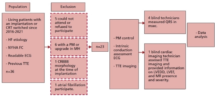
Fig. 1. Study design.
CRBB: complete right bundle block. CRT: cardiac resynchronization therapy. ECG:
electrocardiogram HF: heart failure. LVEDD: left ventricular end-diastolic
diameter. LVEF: left ventricular ejection fraction. MH: medical history. MR:
mitral regurgitation. NYHA: New York Heart Association. PM: pacemaker. TTE:
transthoracic echocardiography
Clinical variables
Clinical data, structural and
functional ECGs were collected at the time of implantation based on each
patient’s medical history and procedure report. In addition, data from
outpatient follow-up (Figure 1) were collected, including a brief
case history on subjective clinical improvement after CRT, current NYHA FC, and
details on the pharmacological treatment.
Device control, records, and surface
ECG measures
Based on each patient’s medical
history, the most recent ECG before the date of CRT implantation was obtained
(“pre-CRT” QRS complex, Figure 1). The day when the patient returned
for follow-up, the device was followed with programmers. The biventricular
stimulation percentage (%BiV) was obtained at the time.
There were two new standard 12-lead ECGs per patient, using a FUKUDA DENSHI
CARDIMAX FX-2111 electrocardiograph gauged at a 25 mm/s paper speed and with a
0.1 mV/mm voltage. The first ECG was performed with the CRT device stimulated
according to usual patient programming (stimulated QRS). To assess intrinsic
ventricular activation (“post-CRT” iQRS), a second
ECG was performed 5 minutes after temporary implantation discontinuation,
programming the device as an off (ODO) or on-demand (VVI) CRT with a HR of 40
bpm.
After recording, the device was
reprogrammed with the patient’s previous setup.
These ECG showed presence or absence
of sinus rhythm. Three electrophysiologists and one electrophysiology-trained
cardiologist were asked to measure the duration of QRS complexes (msec) with the most representative leads, concealing any
data that might identify the patients and ensuring that those measuring QRS
complexes ignored CRT response.
Structural variables (TTE)
The transthoracic echocardiography
(TTE) report closest to, and before the date of CRT implantation was obtained
from the patient’s previous medical history. These reports showed pre-CRT LVEF
data, the left ventricular end-diastolic diameter (pre-CRT LVEDD), and the
presence and extent of mitral valve regurgitation (pre-CRT MR).
On the day of the new ECG, and after
checking the device, each patient had a new TTE performed using ultrasound
General Electric Vivid iQ equipment with a 3.5 Mhz transducer, as recommended by the American Society of
Echocardiography. Images of two and four chambers apical and left parasternal
areas were captured using color Doppler. An echocardiography technician, also
blind to clinical data, read the images, estimated the LVEF using the biplane
Simpson method (post-CRT LVEF), measured the LVEDD, and defined the presence
and severity of mitral regurgitation (post-CRT MR).
CRT response definition
The patient was categorized as a
“clinical responder” and/or “structural responder” based on the most common
criteria to evaluate response to CRT (13,16,29–31):
• A “clinical responder” is someone
with subjective improvement and at least one level improvement (reduction) in
the NYHA functional class as compared to the value before the implantation.
• A “structural responder” is someone
with reduced LV diameters by at least 15% and/or increased LVEF by at least
10%.
• A “hyper-responder” is someone with
reduced LV diameters by 30% and/or normal LVEF.
Statistical analysis
Normal data distribution was checked
using the Anderson-Darling normality test. Continuous variables are presented
as mean ± standard deviation (SD), or median and interquartile range (IQR) of
25-75%, as appropriate. Categorical variables are presented with absolute and
relative frequencies. To evaluate changes in the continuous variable of interest,
iQRS, in response to CRT (pre/post CRT analysis), the
t test or Wilcoxon test for paired data was used, as appropriate. Also,
behavior of ΔiQRS as a continuous variable (post-CRT
QRS – pre-CRT QRS in msec) was compared in responders
vs. non-responders (paired t test or Mann-Whitney test, as appropriate). These
analyses were performed for the clinical-structural (CS) response and
hyper-response across the sample and certain subgroups of patients (female vs.
male, baseline QRS >150 msec vs. <150 msec, females with a baseline QRS >150 msec vs QRS <150 msec, patients with vs without
improved MR). A p-value <0.05 was considered statistically significant. All
graphical analyses used software Graph Pad Prism, version 9.0.
Ethical considerations
The ethical principles set forth in
the Declaration of Helsinki and latest amendments for research in humans were
followed. All patients were called to be informed about the study prior to
enrollment. At the time of evaluation in the clinic, each patient gave their
written informed consent. The study and informed consent provided to the
patients had been previously approved by the Hospital de Clínicas
Ethics Committee.
RESULTS
Study population characteristics
A total number of 23 patients were
enrolled. Clinical, echocardiographic, implant-related, and
electrocardiographic variables are displayed in Table 1.
Table 1. Baseline study population characteristics (pre- CRT)
|
Clinical
variables |
|
|
Female, n
(%) |
12 (52) |
|
Age at
the time of implantation (years), mean+SD |
60±12 |
|
Time
since implantation (months), mean+SD |
43±20 |
|
Non-ischemic
etiology (n, %) |
19 (82) |
|
Medications,
n (%) |
|
|
o
ACEi/ARB |
20 (86) |
|
o
BB |
23 (100) |
|
o
MRA |
18 (78) |
|
o
OMT |
17 (73) |
|
Pre-CRT
NYHA FC, n (%) |
|
|
o
II |
9 (39) |
|
o
III |
14 (61) |
|
Echocardiographic
variables |
|
|
LVEF (%) mean+SD |
26.3±5.7 |
|
LVEDD
(mm), mean+SD |
66.6±12.7 |
|
At least
moderate MR, n (%) |
10 (43) |
|
ECG
variables |
|
|
Duration
of intrinsic QRS (ms), median (IQR 25-75) |
160 (150-170) |
|
Duration
of stimulated QRS (ms), mean+SD
|
119±19 |
|
BiV implantation (%), median (IQR
25-75) |
99 (95-100) |
ACEi:
angiotensin-converting enzyme inhibitors. ARB: angiotensin II receptor
blockers. BB: beta-blockers. BiV: bi ventricular.
CRT: cardiac resynchronization therapy. IQR: interquartile range. LVEDD: left
ventricular end-diastolic diameter. LVEF: left ventricular ejection fraction.
MR: mitral regurgitation. MRA: mineralocorticoid receptor antagonists. NYHA FC:
New York Heart Association functional class. OMT: optimal medical therapy. SD:
standard deviation.
The patients across the sample showed
a similar per-sex distribution; 12 were female (52%). Four (18%) patients had
ischemic heart disease, and 19 (82%) patients had non-ischemic heart disease.
The average age at the time of implantation was 60±12 years, while the age at
the time of follow-up was 64±12 years. The mean follow-up from implantation to
control was 43±20 months, ranging from 7 to 96 months.
A high rate of treatment adherence
was observed among patients; 73% were under simultaneous therapy with
beta-blocker (BB), angiotensin converting enzyme inhibitors (ACEi)/angiotensin II receptor blockers (ARB)/sacubitril-valsartan, and mineralocorticoid receptor
antagonists (MRA). All patients were receiving BB at the time of follow-up.
The duration of the intrinsic QRS
complex prior to CRT was 160 (150-170) msec, and
after the CRT, it was 160 (140-160) msec. Nine (39%) patients showed at least a
10 msec reduction in the iQRS
duration. Maximum reduction was 40 msec and occurred
in three patients. The iQRS increased in eight
patients and remained stable in six.
Analysis of response to CRT
Table 2 shows the
characteristics of CRT responders. Twenty-one (91%) patients in our study were
“clinical responders”. The ΔiQRS in this
group was -4.7±20.4 msec. Structurally and functionally, both LVEDD and
increased LVEF showed enhanced response to CRT during follow-up. LVEDD
decreased from 66.6±12.7 mm pre-CRT to 61.7±12.9 mm post-CRT, with a higher
than 15% reduction in 7 patients (30.4%). LVEF increased from 26.3±5.7% to
41.6±13.4%, with a higher than 10% increase in 16 patients (70%).
Table 2. ECG and clinical response to CRT (pre- and post- CRT analysis)
|
Response parameters |
Pre-CRT |
Post-CRT |
p-value |
|
Structural
variables |
|
|
|
|
LVEF (%) mean+SD |
26.3±5.7 |
41.6±13.4 |
<0.0001 |
|
LVEDD
(mm), mean+SD |
66.6±12.7 |
61.7±12.9 |
<0.0001 |
|
ECG
variables |
|
|
|
|
QRS (ms), median (IQR 25-75) |
160 (150-170) |
160 (140-160) |
0.63 |
CRT: cardiac resynchronization therapy. LVEDD: left ventricular
end-diastolic diameter. LVEF: left ventricular ejection fraction;
Fifteen (65%) patients were
considered “responders” based on both structural and clinical criteria. The
LVEDD was significantly reduced from 64.6±8.7 mm to 59.8±7.5 mm, and the LVEF
increased from a baseline value of 25.5±6.8% to 40.6±9.5% (Table 2). Nine (39%)
patients were “hyper-responders”. All of them had clinical response.
As shown in Table 2, the pre-
and post-CRT analysis of the total number of patients found that the duration
of QRS did not vary significantly following the CRT.
Figure 2 shows a
sample case, with iQRS measurements before and after
the CRT in a “responder”.
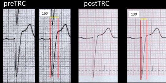
Fig. 2. Reverse electrical remodeling of the left ventricle before and after
CRT. (Images are repeated using calipers and measures.) This patient showed
increased LVEF from 28% to 52%, and decreased LVEDD from 77 to 58 mm.
CRT: cardiac resynchronization
therapy
Subgroup analysis
There was a trend in iQRS duration variation after the CRT in the female
subgroup, though it lacked statistical significance (p=0.056, Figure 3B). Changes in
the duration of iQRS were not higher in those with a
baseline QRS of ≥150 msec (p=0.65, Figure 3C). Upon
analysis of the subgroup of women with an initial QRS ≥150 msec
prior to CRT, significant reduction of the iQRS was
observed (p=0.0195, Figure 3D).
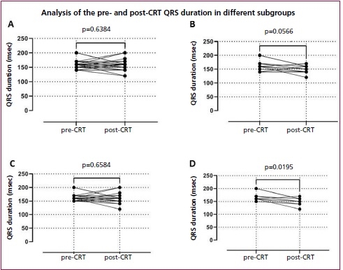
Fig. 3. Differences in iQRS before and after the CRT in the entire population (A),
the female subgroup (B), individuals with an initial QRS ≥150 msec (C), and women with an initial QRS ≥150 msec (D).
CRT: cardiac resynchronization
therapy.
The change in pre- and post-CRT iQRS duration was unrelated to the non-ischemic etiology
(p=0.72) or to age (p=0.78).
The ΔiQRS was
-9.3±20.7 msec among clinical-structural
“responders,” and 11.25±18.9 msec among
non-responders (p=0.027). This difference was slightly larger among
“hyper-responders,” with a ΔiQRS of
-14.44±17.40 msec (p=0.026). There was no significant
association of ΔiQRS with MR improvement (p=0.84). (Figure 4)
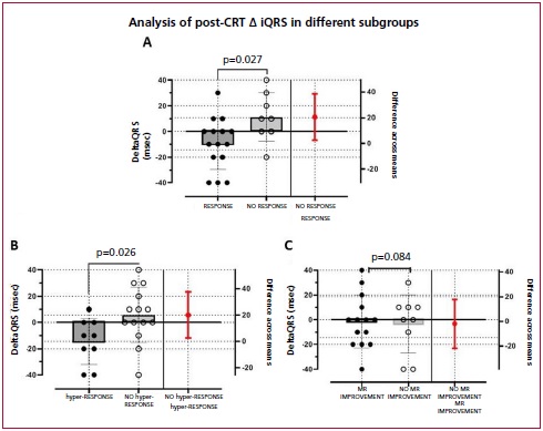
Fig. 4. ΔiQRS charts are shown
by subgroups. A: Clinical-structural response to CRT (p=0.027); B:
Hyper-responders; C: Mitral regurgitation improvement.
CRT: cardiac resynchronization
therapy
DISCUSSION
To the best of our knowledge, this is
the first study to describe reverse electrical remodeling in patients under
local and regional CRT.
After CRT, the duration of iQRS was reduced at least 10 msec
in nine of the patients, though this lacked statistical significance. In
observational studies like ours, QRS reduction was significant and generally
associated with increased LVEF. (21) Subgroup analysis shows that ΔiQRS is related
to clinical-structural response, more markedly in patients having a
“hyper-response” to CRT. Despite sample size limitations, our findings
contribute to the hypothesis that a shorter QRS leads to an improved
intraventricular conduction system, and therefore, reverse LV remodeling.
Further investigation is required to assess this hypothesis.
We showed a tendency towards stronger
electrical remodeling in women with a wider baseline QRS. This is consistent
with traditional response definitions. (1, 31-33)
Mechanisms resulting in reverse electrical
remodeling are not fully known. It might be said that improvements in the size
of chambers favor faster conduction upon myocardium contraction. It has also
been suggested that this may happen as a result of full or partial recovery of
the specialized conduction system itself. (22, 33,34) Another possible CRT-related effect
is reduced cardiac fibrosis. (35) The underlying question also
involves pathophysiological complete LBBB mechanisms and their multiple physiological
and anatomical variants, an electrocardiographic pattern that may lead to
completely interrupted or delayed conduction across the left bundle branch,
which might result in right bundle branch overexpression. (36)
The cutoff point used to define
electrical remodeling is even less consistent. Sebag
et al. prospectively enrolled 85 patients with CRT indication, and evaluated
clinical, echocardiographic, and electrocardiographic variables before and 12
months after the CRT. They found 19 and 18 msec
cutoff points, with a sensitivity and specificity of 86/60% and 84/60%,
respectively, to associate ΔiQRS with the
clinical and echocardiographic response, respectively. Following a multivariate
analysis, ΔiQRS ≥20 msec
was an independent predictor of the echocardiographic response. (26)
We showed a tendency towards a
reduced native QRS relative to improved MR, reaching no significance. The study
by Karaca et al. found that reverse electrical
remodeling is associated with improved MR and geometry of the mitral valve
anatomy. (34) According to them, in addition to
secondary improvement thanks to the cited ventricular remodeling, reversed
papillary muscles dyssynchrony may lead to a major
CRT-related benefit, and both clinical and functional enhancement.
Future prospective investigations are
required to assess the clinical, structural and electrical response in several
specific subpopulations of patients undergoing CRT, in an attempt to better
understand the pathophysiology of its effects. We consider this becomes more
relevant and that it will merit new studies to know if the reverse electric
remodeling is similar or more accentuated in the era of stimulation of the
specific conduction system. (37, 38)
Conflicts of interest
None declared.
(See authors conflicts of interest
forms in the website/ Supplementary material)
 https://creativecommons.org/licenses/by-nc-sa/4.0/
©Revista Argentina de Cardiología
1. Glikson M, Nielsen JC, Kronborg MB, Michowitz Y, Auricchio A, Barbash IM, et al.
2021 ESC Guidelines on cardiac pacing and cardiac resynchronization therapy.
Vol. 42, European Heart Journal. Oxford University Press; 2021. p. 3427–520.
2. McDonagh TA, Metra M, Adamo M, Gardner RS, Baumbach A, Böhm M, et al. Corrigendum to: 2021 ESC Guidelines for the
diagnosis and treatment of acute and chronic heart failure: Developed by the
Task Force for the diagnosis and treatment of acute and chronic heart failure
of the European Society of Cardiology (ESC) With the special contribution of
the Heart Failure Association (HFA) of the ESC. European Heart Journal. 2021
Dec 21;42(48):4901–4901.
3. Mosterd A, Hoes AW.
Clinical epidemiology of heart failure. Heart. 2007 Sep;93(9):1137–46.
4. Dunlay SM, Weston SA,
Jacobsen SJ, Roger VL. Risk factors for heart failure: a population-based
case-control study. Am J Med. 2009 Nov;122(11):1023–8.
5. Conrad N, Judge A, Tran J,
Mohseni H, Hedgecott D, Crespillo AP, et al. Temporal trends and patterns in heart
failure incidence: a population-based study of 4 million individuals. Lancet.
2018;391(10120):572–80.
6. Leclercq C, Hare JM.
Ventricular resynchronization: current state of the art. Circulation. 2004 Jan
27;109(3):296–9.
7. Leclercq C, Kass DA. Retiming the failing heart: principles and current
clinical status of cardiac resynchronization. J Am Coll
Cardiol. 2002 Jan 16;39(2):194–201.
8. Olshansky B, Day JD,
Sullivan RM, Yong P, Galle E, Steinberg JS. Does cardiac resynchronization
therapy provide unrecognized benefit in patients with prolonged PR intervals?
The impact of restoring atrioventricular synchrony: an analysis from the
COMPANION Trial. Heart Rhythm. 2012 Jan;9(1):34–9.
9. Gervais R, Leclercq C, Shankar A, Jacobs S, Eiskjaer
H, Johannessen A, et al. Surface electrocardiogram to
predict outcome in candidates for cardiac resynchronization therapy: a
sub-analysis of the CARE-HF trial. Eur J Heart Fail.
2009 Jul;11(7):699–705.
10. Friedman DJ, Bao H, Spatz ES, Curtis JP, Daubert JP,
Al- Khatib SM. Association Between a Prolonged PR
Interval and Outcomes of Cardiac Resynchronization Therapy: A Report From the
National Cardiovascular Data Registry. Circulation. 2016 Nov 22;134(21):1617–28.
11. Cleland JGF, Daubert JC,
Erdmann E, Freemantle N, Gras D, Kappenberger L, et
al. The effect of cardiac resynchronization on morbidity and mortality in heart
failure. N Engl J Med. 2005 Apr 14;352(15):1539–49.
12. Bristow MR, Saxon LA, Boehmer J, Krueger S, Kass DA, de
Marco T, et al. Cardiac-resynchronization therapy with or without an
implantable defibrillator in advanced chronic heart failure. N Engl J
Med. 2004 May 20;350(21):2140–50.
13. Linde C, Abraham WT, Gold MR, St John Sutton M, Ghio S, Daubert C, et al.
Randomized trial of cardiac resynchronization in mildly symptomatic heart
failure patients and in asymptomatic patients with left ventricular dysfunction
and previous heart failure symptoms. J Am Coll Cardiol. 2008 Dec 2;52(23):1834–43.
14. Moss AJ, Hall WJ, Cannom
DS, Klein H, Brown MW, Daubert JP, et al.
Cardiac-resynchronization therapy for the prevention of heart-failure events. N
Engl J Med. 2009 Oct 1;361(14):1329–38.
15. Tang ASL, Wells GA, Talajic
M, Arnold MO, Sheldon R, Connolly S, et al. Cardiac-Resynchronization Therapy
for Mild-to-Moderate Heart Failure. New England Journal of Medicine. 2010 Dec
16;363(25):2385–95.
16. Daubert JC, Saxon L,
Adamson PB, Auricchio A, Berger RD, Beshai JF, et al. 2012 EHRA/HRS expert consensus statement
on cardiac resynchronization therapy in heart failure: Implant and follow-up
recommendations and management. Heart Rhythm. 2012;9(9):1524–76.
17. Solomon SD, Foster E, Bourgoun
M, Shah A, Viloria E, Brown MW, et al. Effect of cardiac resynchronization
therapy on reverse remodeling and relation to outcome: multicenter automatic
defibrillator implantation trial: cardiac resynchronization therapy.
Circulation. 2010 Sep 7;122(10):985–92.
18. Ypenburg C, van Bommel RJ, Borleffs CJW, Bleeker GB, Boersma E, Schalij MJ, et al. Long-term prognosis after cardiac resynchronization
therapy is related to the extent of left ventricular reverse remodeling at
midterm follow-up. J Am Coll Cardiol.
2009 Feb 10;53(6):483–90.
19. Steffel J, Ruschitzka F. Superresponse to
cardiac resynchronization therapy. Circulation. 2014 Jul 1;130(1):87–90.
20. Ellenbogen KA, Wilkoff BL, Kay GN, Lau CP, Auricchio
A. Clinical Cardiac Pacing, Defibrillation and Resynchronization therapy.
Fifth. ELSEVIER; 2017. 490–522 p.
21. Henrikson CA, Spragg DD, Cheng A, Capps M, Devaughn K, Marine JE, et al.
Evidence for Electrical Remodeling of the Native Conduction System with Cardiac
Resynchronization Therapy.
22. Dizon J, Horn E, Neglia ,
John, Medina N, Garan H. Loss of Left Bundle Branch
Block following Biventricular Pacing Therapy for Heart Failure: Evidence for
Electrical Remodeling? Journal of Interventional Cardiac Electrophysiology. 2004
Feb;10(1):47–50.
23. Qian Z, Qin C, Zou F, Xue S, Wang Y, Zhang X, et al. Complete electrical reverse
remodeling of native conduction after resynchronization therapies. Int J Cardiol. 2022 Jun 15;357:81–7.
24. Stockburger M, Nitardy A, Fateh-Moghadam S,
Krebs A, Celebi O, Karhausen
T, et al. Electrical Remodeling and Cardiac Dimensions in Patients Treated by
Cardiac Resynchronization and Heart Failure Controls.
25. Mischke K, Knackstedt C, Fache K, Reith S, Rana O, Saygili
E, et al. Electrical remodelling in cardiac resynchronization
therapy: decrease in intrinsic QRS duration. Acta Cardiologica. 2011 Apr;66(2):175–80.
26. Sebag FA, Martins RP, Defaye P, Hidden-Lucet F, Mabo P, Daubert JC, et al.
Reverse electrical remodeling by cardiac resynchronization therapy: Prevalence and
clinical impact. Journal of Cardiovascular Electrophysiology. 2012 Nov;23(11):1219–27.
27. Tereshchenko LG, Henrikson CA, Stempniewicz P, Han
L, Berger RD. Antiarrhythmic effect of reverse electrical remodeling associated
with cardiac resynchronization therapy. PACE
- Pacing and Clinical Electrophysiology. 2011 Mar;34(3):357–64.
28. Surawicz B, Childers R,
Deal BJ, Gettes LS, Bailey JJ, Gorgels
A, et al. AHA/ACCF/HRS recommendations for the standardization and
interpretation of the electrocardiogram: part III: intraventricular conduction
disturbances: a scientific statement from the American Heart Association
Electrocardiography and Arrhythmias Committee, Council on Clinical Cardiology;
the American College of Cardiology Foundation; and the Heart Rhythm Society:
endorsed by the International Society for Computerized Electrocardiology.
Circulation. 2009 Mar 17;119(10):e235-40.
29. Hsu JC, Solomon SD, Bourgoun
M, McNitt S, Goldenberg I, Klein H, et al. Predictors
of Super-Response to Cardiac Resynchronization Therapy and Associated
Improvement in Clinical Outcome. J Am Coll Cardiol. 2012 Jun;59(25):2366–73.
30. Gold MR, Rickard J, Daubert
JC, Zimmerman P, Linde C. Redefining the Classifications of Response to Cardiac
Resynchronization Therapy: Results From the REVERSE
Study. JACC: Clinical Electrophysiology. 2021 Jul 1;7(7):871–80.
31. Goldenberg I, Moss AJ, Hall WJ, Foster E,
Goldberger JJ, Santucci P, et al. Predictors of
Response to Cardiac Resynchronization Therapy in the Multicenter Automatic
Defibrillator Implantation Trial With Cardiac Resynchronization Therapy
(MADIT-CRT). Circulation. 2011 Oct 4;124(14):1527–36.
32.
Saxon LA, Bristow MR, Boehmer J, Krueger S, Kass DA, de Marco T, et al. Predictors of sudden cardiac death
and appropriate shock in the Comparison of Medical Therapy, Pacing, and
Defibrillation in Heart Failure (COMPANION) Trial. Circulation 2006;114:2766-72. https://doi.org/10.1161/CIRCULATIONAHA.106.642892
33. Yang XW, Hua W, Wang J, Liu ZM, Ding LG, Chen KP,
et al. Native QRS narrowing reflects electrical reversal and associates with
anatomical reversal in cardiac resynchronization therapy. Journal of
Interventional Cardiac Electrophysiology 2014;41:161–8.
https://doi.org/10.1007/s10840-014-9936-5
34.
Karaca O, Cakal B, Omaygenc MO, Gunes HM, Cakal SD, Kizilirmak F, et al. Native Electrocardiographic QRS
Duration after Cardiac Resynchronization Therapy: The Impact on Clinical
Outcomes and Prognosis. J Cardiac Fail 2016;22:772–80.
https://doi.org/10.1016/j.cardfail.2016.04.001
35. Sunman H, Canpolat U, Yorgun H, Ozkan A, Yalçn MU, Bayrak T, et al.
Association between reverse electrical remodeling and cardiac fibrosis markers
in patients with cardiac resynchronization therapy. Turk Kardiyoloji
Dernegi Arsivi 2018;46:84-91. https://doi.org/10.5543/tkda.2017.80236
36. Tan NY, Witt CM, Oh JK, Cha YM. Left Bundle Branch
Block: Current and Future Perspectives. Circulation: Arrhythmia and
Electrophysiology. Lippincott Williams and Wilkins; 2020. p. 364–77. https://doi.org/10.1161/CIRCEP.119.008239
37. Ponnusamy SS, Arora V, Namboodiri N, Kumar V, Kapoor A, Vijayaraman P. Left
bundle branch pacing: A comprehensive review. J Cardiovasc
Electrophysiol. 2020;31:2462-73.
38.Vijayaraman P, Chung MK, Dandamudi
G, Upadhyay GA, Krishnan K, Crossley G, et al. His
Bundle Pacing. J Am Coll Cardiol
2018;72:927-47. https://doi.org/10.1016/j.jacc.2018.06.017