Evaluación de las placas coronarias por tomografía computarizada multidetector de 16 filas. Correlación con ecografía intravascular
pp 193-199
DOI:
https://doi.org/10.7775/rac.v76i3.2406Palabras clave:
Tomografía computarizada por rayos X, CoronariopatíaResumen
Introducción
El 50% de las muertes de etiología cardiovascular se deben a la enfermedad arteriosclerótica. Estudios recientes demostraron que las placas ateromatosas más propensas al desarrollo de un evento coronario son las que presentan ciertas características particulares, como un centro o core lipídico y remodelación positiva, denominadas placas vulnerables. La determinación temprana de la presencia de este tipo de placas ateroscleróticas tendría un impacto clínico de suma importancia y podría ayudar a prevenir el desarrollo de un síndrome coronario agudo en el futuro.
Objetivo
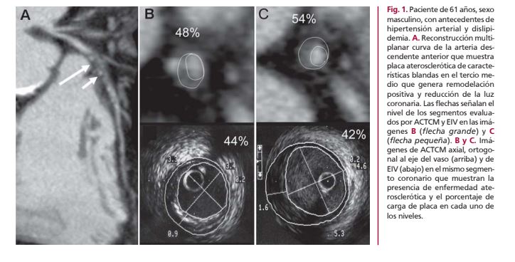
Determinar la certeza diagnóstica de la angiografía coronaria por tomografía computarizada multidetector de 16 filas (16-ACTCM) en la identificación, la caracterización y la cuantificación de las lesiones arterioscleróticas coronarias en comparación con la ecografía intravascular (EIV).
Material y métodos
Se estudiaron 45 pacientes con indicación de angiografía coronaria con 16-ACTCM y EIV. En cada segmento coronario se analizaron la carga de placa y las características de la placa aterosclerótica, que se clasificó en blanda, fibrosa y cálcica. La certeza diagnóstica de la 16-ACTCM para determinar la carga de placa y la identificación de placas coronarias se calculó con el método exacto binomial. Sobre la base de un análisis con curvas ROC se determinó el punto de corte para cada tipo de placa, así como la densidad media y la desviación estándar en unidades Hounsfield [UH]). Se evaluó además la certeza diagnóstica de la ACTCM para el diagnóstico de estenosis coronaria ≥ 50%.
Resultados
Para la detección de carga de placa, la sensibilidad fue del 96,2% y la especificidad fue del 81,9%. Para la detección de placas blandas, fibrosas y calcificadas, la sensibilidad y la especificidad fueron del 94,6% y 92,62%, del 94,9% y 98,5% y del 93,2% y 95,1%, respectivamente. Con un valor de corte de 85 UH, la 16-ACTCM diferenció correctamente el 86% de las placas blandas y fibrosas, con un área ROC = 0,96; con un valor de corte de 196,7 UH diferenció correctamente el 93% de las placas calcificadas y no calcificadas, con un área ROC = 0,98. Para la detección de estenosis coronaria, la sensibilidad fue del 81,58% y la especificidad fue del 93,8%.
Conclusión
La 16-ACTCM es una modalidad diagnóstica no invasiva promisoria para el estudio de pacientes con enfermedad coronaria, ya que permite la detección y la caracterización de los diferentes tipos de placa.
Descargas
Publicado
Número
Sección
Licencia
Derechos de autor 2025 Revista Argentina de Cardiología

Esta obra está bajo una licencia internacional Creative Commons Atribución-NoComercial-CompartirIgual 4.0.