
Mieloperoxidasa como indicador de estrés oxidativo en síndrome metabólico
pp 514-518
DOI:
https://doi.org/10.7775/rac.v84i6.1270Keywords:
Myeloperoxidase, Oxidative Stress, Metabolic Syndrome, Insulin Resistance, Cardiovascular RiskAbstract
Background: Increased myeloperoxidase (MPO) activity would be the link between the rise of the inflammatory response and oxidative stress (OS) in metabolic syndrome (MS).
Objective: The aim of this study was to determine the enzymatic activity of MPO associated with OS in animals with MS and establish their relationship with probable cardiovascular injury.
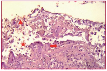
Methods: Male Wistar rats were divided into two groups: Group A, control (n=12) and Group B, induced MS (n=12). Metabolic syndrome was produced by 6-week administration of 10% fructose diluted in the drinking water. Insulin (μU/ml), glucose (mg/dl), lipid panel (mg/dl), HOMA (homeostatic model assessment), MPO (IU/ml) and superoxide dismutase (SOD) activity (U/ml) were measured. Light microscopy was used for the histological study of the heart and thoracic aorta.
Results: Group B showed significantly increased levels of plasma glucose (176±17.3 mg/dl), insulin (29.5±4.52 μU/ml), HOMA(11±1.3), total cholesterol (133±9.6 mg/dl) and triglycerides (75±12.9 mg/dl) compared with Group A: plasma glucose (115±1.1
mg/dl), insulin (4±0.82 μU/ml), HOMA (3±0.38), total cholesterol (69.7±1.6 mg/dl) and triglycerides (46.2±6 mg/dl), (p<0.001 for all variables). A significant decrease in HDL (28.3±1.14 mg/dl) in Group B vs. Group A (61±1.0 mg/dl) (p<0.001) validated the experimental MS model. Myeloperoxidase activity increased significantly in Group B (181.3±15.7 IU/ml) vs. Group A (116.07±4.2 IU/ml) (p<0.001). A similar behavior was seen with SOD antioxidant activity in Group B (181±6 U/ml) vs. Group A (138±3.6 U/ml) (p<0.01). Light microscopy of the heart and thoracic aorta revealed histopathological changes in animals with induced MS.
Conclusion: Increased MPO and SOD in Group B would indicate the presence of OS in MS, with consequences at the vascular level.
Downloads
Published
Issue
Section
License
Copyright (c) 2025 Argentine Journal of Cardiology

This work is licensed under a Creative Commons Attribution-NonCommercial-ShareAlike 4.0 International License.


