
Right Ventricle: Echocardiographic Evaluation of Pressure and Volume Overload
pp 556-562
DOI:
https://doi.org/10.7775/rac.es.v84.i6.9467Keywords:
Echocardiography, Two-dimensional Strain, Pulmonary Hypertension, Right VentricleAbstract
Background: New echocardiographic techniques have renewed the interest in the assessment of right ventricular function; however, there are still not clearly defined reference values or large-scale analyses on the behavior of this chamber when submitted to pressure (RVPO) or volume (RVVO) overload.
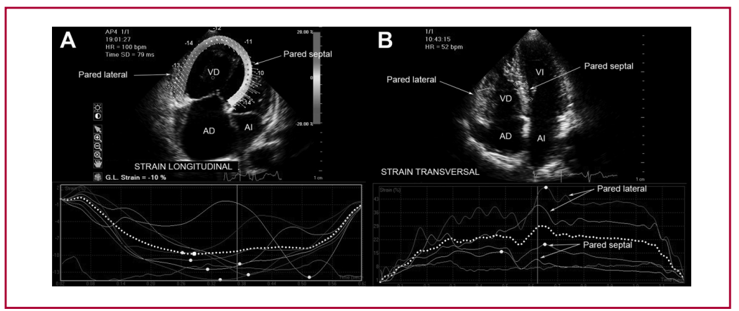
Objective: The aim of this study was to analyze echocardiographic abnormalities and right ventricular longitudinal and transverse strain in patients with RVPO and RVVO and in healthy subjects (CTRL).
Methods: Fifty CTRL subjects (mean age 47±18 years, 31 females), 50 RVPO patients (mean age 48±12 years, 32 females) and 30 RVVO patients (mean age 43±14 years, 19 females) were included in the study. Left ventricular, left atrial, right ventricular and right atrial dimensions, and left and right ventricular function were analyzed. Two-dimensional left and right ventricular longitudinal strain and right ventricular transverse strain were evaluated. Analysis of variance was used to compare means and standard deviations.
Results: Demographic data, left ventricular ejection fraction and adjusted left atrial volume showed no significant differences among groups. Diameters, right ventricular wall thickness, tricuspid regurgitation gradient and adjusted right atrial volume were higher in RVPO patients. Right ventricular function parameters were decreased in RVPO and normal in RVVO patients. The right ventricular
lateral transverse strain was increased both in RVPO and RVVO.
Conclusions: Right ventricular echocardiographic parameters and strain showed signs of remodeling, both in RVPO and RVVO,and decreased systolic function in RVPO. Increased right ventricular transverse strain would probably be due to remodeling, and decreased longitudinal strain to systolic dysfunction. Decreased left ventricular longitudinal strain may indicate early biventricular dysfunction.
Downloads
Published
Issue
Section
License
Copyright (c) 2025 Argentine Journal of Cardiology

This work is licensed under a Creative Commons Attribution-NonCommercial-ShareAlike 4.0 International License.


