
Ischemia-Reperfusion Experimental Models Electrocardiographic and Morphologic Findings
pp 141-150
DOI:
https://doi.org/10.7775/rac.v62i2.3335Keywords:
Ischemia reperfusion, Chloricromene, Myocardial damage, Oxidative stressAbstract
Background and objectives
Varios experimental studies on prevention and treatment of myocardial damage due to ischemia-reperfusion phenomena as well as the pathogenic mechanisms involved have been published. The aim of this study is to describe the electrocardiographic and morphologic findings in rabbit hearts submitted to experimental ischemia and reperfusion.
Methods Forty rabbits were divided into 4 groups: group 1: occlusion of the left anterior descending artery; group2: occlusion of the left circunflex artery; group 3: occlusion of circunflex" artery + infusion of AD6, 6.4 pg/kg/min, and group 4: occlusion of circunflex artery +saline infusion. Each group consisted of 10 New Zealand rabbits. After 50 minutes of occlusion, the artery was reopened for 20 minutes. Then, biopsy specimens of the myocardium at risk and of the perfused tissue were obtained. Electrocardiograms were recorded throughout the experiment. In 3 animals of group 1, a marked widening of the QRS complex was observed whilst arrhythmia during reperfusion occurred in 4. No animals in this group died. The electrocardiograms of rabbits in group 2 showed ST-segment shifting, most frequently a 3-8 mm elevation, during ischemia and reperfusion. One animal presented a non-sustained ventricular tachycardia and 5 died due to malignant sustained ventricular tachycardia and/or ventricular fibrillation. In group 3 only transient ST-segment elevation was observed in 3 animals. There were significant differences between group 3 and 4 regarding ST-segment changes during ischemia and reperfusion (p <0.05). In group 3, reversal of ventricular fibillation was observed in 2 rabbits after increasing the infusion rate of AD6 to 10.7 mg/kg/min; ST-segment elevation also retrograded. Two animals in this group and 5 in group 4 died. After sacrifice, the coronary arteries were injected with either triphenyltetrazolium or methylene blue to determine the area at risk, which corresponded to 33 ± 18% of the tissue in group 1, to64 ± 15% in group 2, to 64.7 ± 6 % in group 3 and to 62.6±6% in group 4. Results Wide areas of focal subendocardial necrosis, contraction bands and focal hemorrhage, affecting less than 10% and 68.4±8.9°í of the left ventricle, were observed in group 1 and group 2, respectively. In group 3, 41.5 ± 3.1 % of the left ventricle was damaged (sub-endocardial necrosis). In the 5 surviving rabbits of group 4 large areas* of wavy fibers which involved 64.7 ± 6% of the left ventricle, were detected. The 5 animals that died earlier due to ventricular fibrillation only showed focal or confluent areas of contraction bands. Ultrastrucurally, subsarcolemmal bubbles, sarcolemal disruption, wide edematous areas, contraction bands, scarce glycogen deposits, marked mithocontrial edema with disruption of the internal and external membranes, and dense deposits were observed in the necrotic tissue.In the ischemic areas, loss of glycogen, chromatin condensation, intermyofibrillar edema and of the sarcoplasmic reticulum were found. Conclusions The ultrastructurally observed lesions were similar all 4 groups, but the area involved was appeared to be important only when the circunflex artery was occluded (group 1 versus group 2). This experimental model allowed the evaluation of the cardioprotective effect of AD6 (group 3 versus group 4). The occlusion of the circunflex artery in the rabbit seems to be an adequate experimental model for ischemia and reperfusion.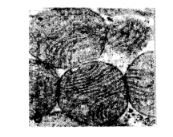
Downloads
Published
2026-03-31
Issue
Section
ORIGINAL ARTICLES
License
Copyright (c) 2026 Argentine Journal of Cardiology

This work is licensed under a Creative Commons Attribution-NonCommercial-ShareAlike 4.0 International License.


