New Imaging Technologies for the Diagnosis and Treatment of Complex Aortic Aneurysms
pp. 522-527
DOI:
https://doi.org/10.7775/rac.es.v88.i6.19346Keywords:
Aortic Aneurysm, Abdominal, Endovascular Procedures, Intraoperative Images, CBCT, Image FusionAbstract
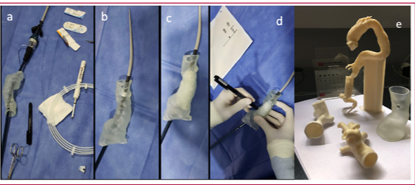
Imaging technology plays a key guiding role in the endovascular treatment of aortic aneurysms, especially in cases of complex thoracoabdominal aortic aneurysms. The operating rooms combine high quality imaging with a sterile and functional environment, allowing for better outcomes, a reduction of the contrast agent to be used and of patients’ and operators’ exposure to radiation. The aim of this presentation is to describe the current use of imaging technology, combining intraoperative cone beam computed tomography (CBCT) and computed tomography angiography (CTA), image “fusion” and intravascular ultrasound, to guide the procedures and thus improve the intraoperative success rate and reduce the need for reinterventions. Also, a procedure to create custom 3D templates with high-definition images of the patient’s arterial anatomy is described, which serve as specific guides to develop fenestrated stent-grafts in the operating room. These custom fenestration templates could increase the number of patients with complex aneurysms treated with a minimally invasive approach.
Downloads
Published
Issue
Section
License
Copyright (c) 2025 Argentine Journal of Cardiology

This work is licensed under a Creative Commons Attribution-NonCommercial-ShareAlike 4.0 International License.