
Usefulness of Multi-Detector Row Computed Tomography Angiography in Aortic Diseases
pp 292-299
DOI:
https://doi.org/10.7775/rac.v79i3.2274Keywords:
Diagnostic Imaging, Aorta, Tomography, Aneurysm, DissectionAbstract
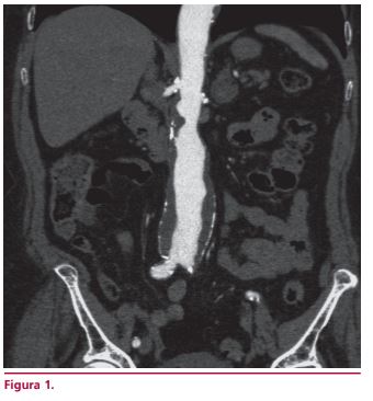
The goal of the present review is to demonstrate the usefulness of multi-detector row computed tomography (MDCT) angiography in the evaluation of diseases of the aorta. The high morbidity and mortality associated with this condition requires a rapid diagnostic tool with diagnostic accuracy at the moment of evaluating patients with known or clinically suspected disease of the aorta. The non invasive nature of and the rapid evaluation provided by MDCT angiography are the main advantages of the method that is widely accepted by the patients. MDCT angiography is the reference-standard method for the evaluation of aneurysms of the aorta, describing its location in the spatial planes, extension, diameters and characteristics of the aortic wall. The clinical presentation of the acute aortic syndromes - aortic dissection, intramural hematoma and penetrating aortic ulcer - is similar. MDCT angiography is a diagnostic tool with the greatest efficacy to confirm or rule out aortic lesions. The multiple visualization techniques and the possibility of multiplanar and three-dimensional reconstructions make it easy to choose between surgical or endovascular treatment.
Downloads
Published
Issue
Section
License
Copyright (c) 2025 Argentine Journal of Cardiology

This work is licensed under a Creative Commons Attribution-NonCommercial-ShareAlike 4.0 International License.


