Vascular Expression of Proinflammatory Transcription Factors in a Model of Metabolic Syndrome
pp 36-41
DOI:
https://doi.org/10.7775/rac.v75i1.2535Keywords:
Remodeling, Metabolic Syndrome, Inflammation, Transcription genesAbstract
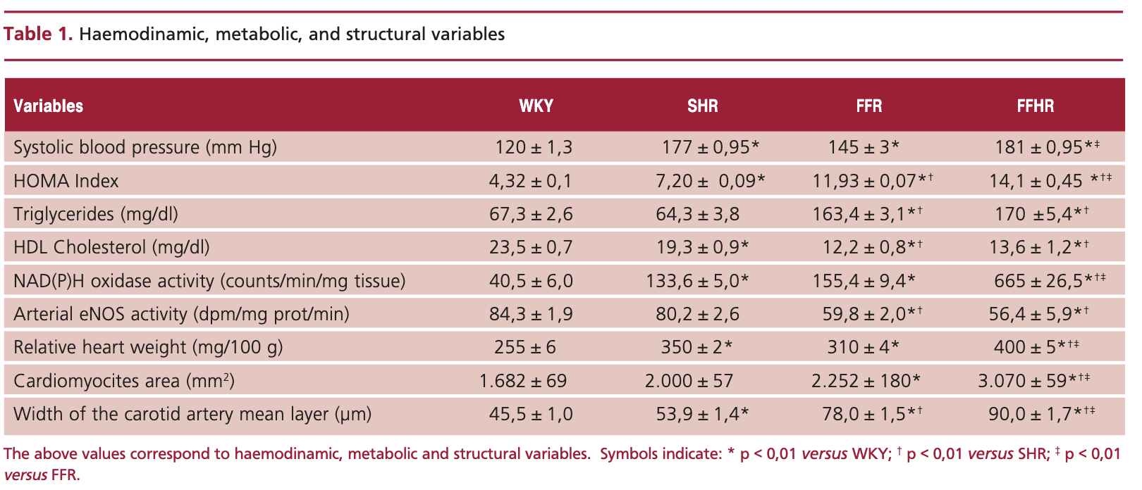
With the goal of assessing the expression of redox-sensitive transcription factors in the arteries of an experimental model of hypertension associated to metabolic syndrome (FFHR), we studied Wistar Kyoto rats (WKY) and spontaneously hypertensive 30-day old male rats (SHR), which were randomly distributed in 4 groups (n=8 in each group). Group 1: WKY (control rats), Group 2: FFR: rats that received 10% W/V fructose in drinking water during a 10-week period, Group 3: SHR rats and Group 4: FFHR: 2+3, i.e., SHR rats treated like Group 2. Groups FFR and FFHR had HOMA (homeostasis assessment model) index and area under the curve (AUC) values in the glucose tolerance test, that were characteristic of insulin resistance. They also showed significant differences in plasma triglyceride and HDL cholesterol levels compared to controls, and increased their systolic blood pressure. Oxidative stress, as assessed by NAD(P)H oxidase activity and TBARS (thiobarbituric acid reactive substances) plasma concentration were significantly higher in FFR and FFHR groups, whereas in these same groups eNOS activity decreased markedly. Relative cardiac weight increased in FFR and FFHR groups, with a larger myocites area in the left ventricular free wall. Sections of the left carotid artery exhibited eutrophic growth of the media layer in FFHR. Average optical density for anti-c-fos, anti-NF-κB and anti-VCAM-1 antibodies was greater in resistance renal arteries and in the carotid artery of FFHR and FFR groups. The data confirm the findings of the pathological experimental model and suggest that oxidative stress and the subsequent activation of genes that participate in the inflammatory process are actively involved in the development of vascular remodeling.
Downloads
Published
Issue
Section
License
Copyright (c) 2026 Argentine Journal of Cardiology

This work is licensed under a Creative Commons Attribution-NonCommercial-ShareAlike 4.0 International License.