
Chagas' disease: A 12 year follow-up in an urban area
pp 205-216
DOI:
https://doi.org/10.7775/rac.v60i2.3287Abstract
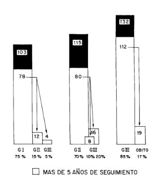
Three hundred fifty patients suffering from chronic Chagas' disease, 120 with a follow-up of 12 years and 270 with more than 5 years follow-up were studied. As suggested by the Committee of Chagas' Disease of the Argentine Society of Cardiology, the 350 patients were classified in three groups, according to the degree of myocardial involvement, namely: group I, n = 103; group II, US, and group m, 132 patients. In group I, patients were in GI functional capacity at the begining of the study, however, 16 (20 %) out of 78 (75 %) with more of 5 years of follow-up presented a progressive myocardial damage, passing 12 to the group II because of electrocardiographic disturbances and 3 to the group III, because of it and of cardiomegaly. In group II, 80/U5 (70 %) had more than 5 years follow-up; 8 (10 %) of them presented a worsening of the conduction disturbances; 16 (20%) passed to the group III because of cardiomegaly. In group III, 112/132 (85 %) presented more than 5 years follow-up; 19 (24 %) suffered from an "expected death". Of note, only 19 patients belonging to the group III (severe myocardial damage) died (19/ 350) representing a 5 % of global mortality. The three groups showed high values of antilaminin in their sera, but the highest values were observed in some patients of group III. These patients suffered also from apical ventricular aneurysms and severe arrhythmias. In 30 patients an endomyocardial biopsy was obtained; mild to moderate hypertrophy of myocytes as well as variable degrees of interstitial fibrosis were observed. Of note, extensive infiltrates consisting of macro phages and mononuclear cells ("true lymphocytes", when were stained with specific monoclonal antigens) were observed. The majority of lymphocytes belonged to the T-type. The cellular infiltration was associated with necrotic and degenerative myocardial lesions. From the ultrastructural point of view, the most striking alteration was represented by the thickening of the basal membrane of myocytes and vascular smooth muscle cells. Immunohistochemical staining for laminin, revealed clearly positive staining in the thickening basement membranes of all patients studied. Basement membrane- like material present within the lumina of T-tubules of myocytes also showed a positive reaction. The implication of the deposit of antilaminin at that site deserves further studies.
Downloads
Published
Issue
Section
License
Copyright (c) 2026 Argentine Journal of Cardiology

This work is licensed under a Creative Commons Attribution-NonCommercial-ShareAlike 4.0 International License.


