
Computed Tomography in the Diagnosis of Aortic Dissection
pp 368-373
DOI:
https://doi.org/10.7775/rac.v60i4.3314Abstract
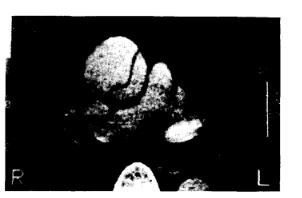
Computed axial tomography is a well established method for the initial evaluation of patients suspected of having aortic dissection. It has the advantage of being non-invasive, quick, and interpretation is easily accesible to clinicians and surgeons. The disadvantages are that it requires the use of radiological contrast material, it does not allow for evaluation of aortic regurgitation nor identification of the exact site of the tear, the reentry site and the possible involvement of important aortic branches. 38 consecutive patients with suspected aortic dissection on clinical grownds are presented. All patients were submitted to a CT scan and evaluated by another diagnostic procedure and/or surgical intervention. A positive diagnostic was made in 17 patients by at least two methods (13 type A and 4 type B). In 14 of these cases the CT scan showed direct signs of dissection: flap, double lumen and/or displacement of intimal calcification. In the three remaining cases only significant dilatation of the aorta was present. The most frequent diagnostic pitfalls were the use of inappropiate tomographye technique or missinterpelation of anatomical structures.
Downloads
Published
Issue
Section
License
Copyright (c) 2026 Argentine Journal of Cardiology

This work is licensed under a Creative Commons Attribution-NonCommercial-ShareAlike 4.0 International License.


