
Spontaneous Closure of Ventricular Septal Defects. An Ignored Cause of Atrioventricular and Intraventricular Conduction Disturbances
pp 165-174
DOI:
https://doi.org/10.7775/rac.v64i2.3378Keywords:
Intraventricular block, Atrioventricular block, Congenital heart disease, Ventricular septal defect, Spontaneous closure of a ventricular septal defect, Conducting system, Sudden deathAbstract
Background The perimembranous ventricular septal defect is the most common form of this congenital abnormality. More than 50% of these defects undergo spontaneous closure in the first year of age. Two mechanisms have been recognized for the spontaneous closure: fibrous proliferation and adherence of the septal leaflet of the tricuspid valve. The branching segment of the His bundle lies in the posterior and inferior edge of the defect. The fibrous tissue developed in the process of spontaneous closure of the septal defect may involve the conducting tissue provoking different types of intraventricular and/or atrioventricular conduction disturbances. This is a commonly ignored cause of conduction disturbances and it may be well over-looked if not advised of this possibility. Material and method Ten patients (5 men and 5 women between 6 and 42 years old) showing intraventricular and/or atrioventricular conduction disturbances with normal or apparently normal hearts were studied. None of the patients had murmurs. The X ray's and the size and width of atria and ventricles were normal. All cases showed evidences indicating that the conduction disturbances may be related to the spontaneous closure of a ventricular septal defect. The observed conduction disturbances were: isolated right bundle branch block in 4 (two of these with 1st degree atrioventricular block), right bundle branch block with left anterior hemiblock in 2 (one of them with second degree atrioventricular block), left bundle branch block in 1 and isolated left anterior hemiblock in 4. Results Spontaneous closure was documented by histopathologic study in 1 case, angiography in 2 and 2-D echocardiography in the other 7 cases. Angiography disclosed a round calcification of about 8 mm diameter at the site of closure in one case and in the other a small infracristal defect that closed spontaneously 5 years later with the formation of apseudoaneurysm as shown by an eco-Doppler study. In the other 7 patients the diagnosis of spontaneous closure was suspected based on echocardiographic findigs. Subxiphoideal recordings showed adherence of the septal leaflet of the tricuspid valve and/or marked distortion of upper part of the ventricular septum. In the case with histological study of the conducting system there were clear cut evidences of the causal relationship between the spontaneous closure and its pathologic changes with the conduction disturbances: right bundle branch block, left anterior hemiblock and second degree atrioventricular block. Conclusions In conclusions, in every child, teenager or youngster showing a conduction disturbance with a normal or apparently normal heart, we must search for echocardiographic evidences of spontaneous closure of a ventricular septal defect. Obviously, any finding suggesting this possibility has important diagnostic and prognostic implications.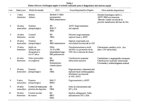
Downloads
Published
2026-03-12
Issue
Section
ORIGINAL ARTICLES
License
Copyright (c) 2026 Argentine Journal of Cardiology

This work is licensed under a Creative Commons Attribution-NonCommercial-ShareAlike 4.0 International License.


