
Macrophage Infiltration in Primary Coronary Plaque Tissues is Associated with Restenosis after Percutaneous Coronary Intervention
pp 199-207
DOI:
https://doi.org/10.7775/rac.v64i2.3381Keywords:
Macrophages, Restenosis, AtherectomyAbstract
Background
Restenosis remains the major limitation of percutaneous coronary revascularization. Macrophages release matrix metalloproteinases and growth factors which may induce smooth muscle cell migration and proliferation. We tested the hypothesis that: 1) primary lesions that develop restenosis after coronary atherectomy have more macrophagesthan primary lesions that do not develop restenosis; and 2) angiographic restenosis is related to the macrophage content of the primary lesion.
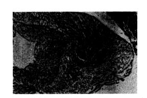
Method and resultsFifty atherectomy specimens from the culprit lesion of patients with unstable angina were examined. Hematoxylin and eosin, trichrome and immunostainipg with anti-human macrophage mono-clonal antibody (KP-1) were performed. Follow-up angiogiaphy was performed at 16±2 weeks. Restenosis, defined as > 50% diameter stenosis(quantified coronary arteriography) was present in30 patients. The other 20 patients (<50% stenosis) were the control group. Coronary risk factors where similar in both groups. Using computer aided planimetry, the total tissue area examined was 5.03±0.4 mm' for atherectomy specimens from patients with restenosis and 5.26 ±0.72 mm' for patients with no restenosis (p= NS).The percentage of macrophage rich areas was significantly larger in plaque tis-sue from patients with restenosis (19.7 ±3%) than in tissue from patients with no restenosis (10.3±2%) (p = 0.006). Linear regression analysis showed that percent diameter stenosis, late loss in MLD and the relative late loss index at FUA were significantly related to the macrophage content at the time of DCA (p<0.01).
Conclusion
Macrophage rich areas are significantly larger in primary lesions from patients that develop restenosis than in those patients that do not develop restenosis.The increased macrophage content suggests that these inflammatory cells are actively involved in the complex process of restenosis after percutaneous coronary revascularization.
Downloads
Published
Issue
Section
License
Copyright (c) 2026 Argentine Journal of Cardiology

This work is licensed under a Creative Commons Attribution-NonCommercial-ShareAlike 4.0 International License.


