Anatomical investigation of the cardiac apex
pp. 118-123
DOI:
https://doi.org/10.7775/rac.es.v90.i2.20498Keywords:
Anatomy and histology - Myocardium - Heart Ventricles - Ventricular FunctionAbstract
Objective: Understanding cardiac anatomy is the key to solve unknown issues about its function. The continuous and helical myocardial structure plays a fundamental role in its torsion-detorsion motions. Does the apex, a constitutive part of the ventricle, have relevance in cardiac dynamics or is it simply a cul-de-sac? The aim of this study was to answer this question.
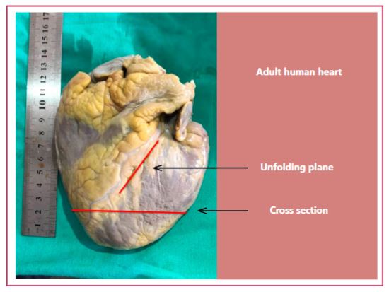
Methods: Four young bovine and four human hearts (two embryos and two adults) were used for the anatomo-histological studies. Two procedures were carried out for this investigation: a) the continuous myocardium unfolding to observe the fiber arrangement at the tip of the left ventricle, called the apical zone; and b) horizontal and longitudinal sections to study the structure of the apex. The horizontal sections were performed between the middle 2/3 and the apex, and the longitudinal ones, sectioning left ventricular apex, with an apex-base orientation.
Results: In all the human and bovine hearts studied we found that the apex corresponds only to the left ventricle, where the twist of the descending segment is located, in the ascending continuity of the myocardium. The apical cul-de-sac has practically no muscular plane at its end. It is internally lined by the endocardium and externally by the epicardium. The muscular plane has only 10% thickness of the adjacent myocardium, a structural concept confirmed by transillumination.
Conclusions: The apex is a cul-de-sac practically devoid of muscle, in which the endocardium and epicardium are attached, but which performs the functions of supporting intraventricular pressures and being a constitutive part of the torsion and detorsion motions.
How to cite this article:
Trainini J, Beraudo M, Wernicke M, Carreras Costa F, Trainini A, Mora Llabata V, et al. Anatomical Investigation of the Cardiac Apex. Rev Argent Cardiol 2022;90:118-23. http://dx.doi.org/10.7775/rac.v90.i2.20498