
Lung Ultrasound in Cardiology: A Window to Pulmonary Edema
pp .463-468
DOI:
https://doi.org/10.7775/rac.es.v87.i6.16767Abstract
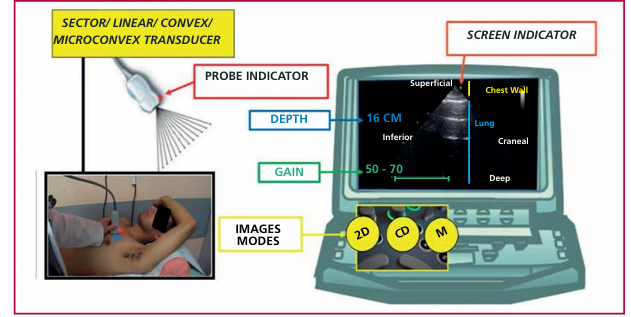
Lung ultrasound is a novel diagnostic tool that has revolutionized the way of performing ultrasonography and cardiology based on the interpretation of artifacts and modifying the diagnostic and therapeutic approach of patients with heart diseases. This technique can be performed by the cardiologist with the transducer and screen adjustments used in echocardiography and should recognize four ultrasound patterns: aerated/dry lung, wet or interstitial lung, pleural effusion and consolidation. The interstitial pattern is the basis of the applications of this technique in cardiology, which is summarized in seven clinical settings: differential diagnosis of dyspnea, heart failure syndrome, identification of acute respiratory distress, extreme situations as high altitude pulmonary edema, breath-hold diving and ironman, dialysis patients, acute coronary syndrome and alveolar-capillary stress echo. Developing skills in cardiopulmonary ultrasound should be mandatory and not optional for cardiologists.
Downloads
Published
Issue
Section
License
Copyright (c) 2019 Argentine Journal of Cardiology

This work is licensed under a Creative Commons Attribution-NonCommercial-ShareAlike 4.0 International License.


