Artificial Intelligence–Driven Analysis of Infrared Thermography for Noninvasive Diagnosis of Low Cardiac Output in Critically Ill Patients
pp. 283-288
DOI:
https://doi.org/10.7775/rac.es.v94.i4.21036Keywords:
Thermography, Artificial intelligence, Hemodynamics, Critical care, Shock, Heart failureAbstract
Introduction: Cutaneous blood flow plays a key role in thermoregulation and is altered in states of hemodynamic shock. Infrared thermography offers a non-invasive method to map temperature and analyze its behavior in patients with hemodynamic
instability.
Objective: To evaluate the thermal behavior of the face in patients under invasive hemodynamic monitoring using an infrared camera. Secondarily, to determine the diagnostic value of this method using artificial intelligence.
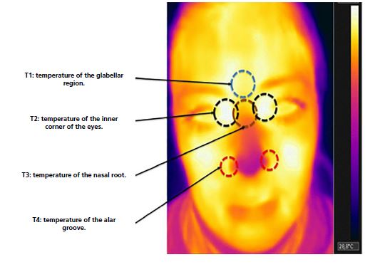
Methods: A cross-sectional study was conducted in critically ill patients monitored with a pulmonary artery catheter. Facial
thermal images were obtained with an infrared camera simultaneously with invasive hemodynamic measurements. Six predefined
facial regions were analyzed, and isolated temperatures, averages, and thermal gradients were calculated. Data were processed using machine learning to predict a cardiac index <2.5 L/min/m².
Results: Thirty-five monitoring sessions were performed in 18 patients (age 60±14 years, 70 % male). Sixty percent presentedwith shock and 40 % were postoperative cardiac patients; 54 % were receiving ≥2 vasoactive drugs. Most facial thermal analyses showed a significant correlation with cardiac index (strongest correlation: thermal gradient, r = –0.45; p = 0.010). The neural network model achieved an area under the ROC curve of 0.75 for predicting low cardiac output.
Conclusions: In this proof-of-concept study, facial thermography identified regions and gradients that significantly correlated
with the cardiac index. Its integration with artificial intelligence exhibited a promising biological signal for the detection of low
cardiac output, emerging as an alternative to be explored for non-invasive hemodynamic monitoring in the critical care setting.
Downloads
Published
Issue
Section
License
Copyright (c) 2026 Argentine Journal of Cardiology

This work is licensed under a Creative Commons Attribution-NonCommercial-ShareAlike 4.0 International License.