
Relation Between Transmural flow and Passive Elastic Stiffness of the Left Ventricle
pp 407-414
DOI:
https://doi.org/10.7775/rac.v62i4.3539Keywords:
Diastolic properties, Passive elastic stiffness, Compliance, Transmural flow, DopplerAbstract
Background
Doppler transmural flow with a predominance of atrial filling (A wave) is regarded as being the consequence of a delayed relaxation while that with a predominance of rapid filling (E wave) would be due to an increased ventricular stiffness.The degree of alteration of the passive elastic stiffness of the left ventricle for these two patterns of transmural flow has not been defined. The purpose of this study was to analize the relationship between transmural flow registered by Doppler and passive elastic stiffness of the left ventricle.
Metbods
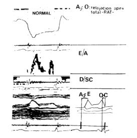
Forty-eight subjects were evaluated, 17 women and 31men (average age 47 ± 12 years). They underwent the following studies: phonocardiogram, apexcardiogram, 2-D echo and transmural flow Doppler. The total apexcardiographic relaxation level was determined (TAR)(A2-O), the diameter of diastole of the left ventricle according to body surface area (D/BSA), peak speed of E and A waves and the E/A relationship were determined.The end diastolic pressure (EDP) was determined based on Palomo's regression equation (21.6 xQ-C/A2-E)+ 1.1. Passive elastic stiffness was evaluated according to two parameters in which volume was replaced by D/BSA:a) the constant k of elastic stiffness (Gaasch), k = (InEDP-InO.43)/(D/BSA); b) the index of chamber stiffness of the left ventricle at the end of diastole(dP/dD).The population was divided into three groups according to the features of the transmural flow: 1) control group(N); 2) patternI (PI) according to Appleton, subjects presenting concentric hypertrophy of the left ventricle; and 3) pattern11 (PII) subjects with dilated cardiomyopathy.
Results
PH presented a largerD/BSA(42 ± 03 cm/m2) (p <0.05) compared to N and PI (2.8±03cm/m2 and 2.6 ± 0.5 cm/m2). EDP was 8 ± I mm llg;12 ±4 mmlg and 18 ± 7 mmHg for N, PI and PH respectively and k was 1 ± 0.1;12 ± 02; and 09 ± 0.1.dP/dD was greater in PH (17 ± 7.5 mmHg/cm/m2) and PI (12 ± 4.5 mmHg/cm/m2) than in N (82 ± 1.7 mmHg/cm/m2)(p < 0.05). TAR was higher in PH (100 ± 30 m sec) and PI (99 ±34 msec) than in N (75 ± 16 m sec) (p < 0.05). E/A was 1.7 ±03(N); 0.7 ±02(PI); and19 ±03(PH). For PI and PH the correlation between kA was -0.52 (p < 0.001) and thec orrelation be DP and E/A was 0.75 (p <00001).
Conclusions
1) When there is abnormal relaxation, the pattern of transmural flow E/A has an inverse relation with the k constant of stiffness. 2) dP/dD is increased for PI and PH, being more altered in the case or the latter group. 3) PH presents a correlation with° less stiff ventricle but the high EDP causes it to operate within the higher portion of aP-D curve thus causing a greater stiffness of the chamber for the said level of EDP. 4) The correlation between EDP and E/A suggests that for PH the decrease of the atrium contribution relies more on the level of EDP than of the passive elastic properties of the left ventricle.
Downloads
Published
Issue
Section
License
Copyright (c) 2026 Argentine Journal of Cardiology

This work is licensed under a Creative Commons Attribution-NonCommercial-ShareAlike 4.0 International License.


