Functional Behavior of Intermediate Coronary Stenoses. Assessment by Intracoronary Doppler, Digital Angiography and Tomographic Stress Perfusion Imaging
pp 139-149
DOI:
https://doi.org/10.7775/rac.v66i2.3482Keywords:
Intracoronary Doppler, Angiography, Myocardial perfusion, Stenoses, Coronary flow reserveAbstract
Objectives
There are significant number of anatomic and pathophysiologic factors that influence the functional behavior of intermediate coronary stenoses, leading to a difficult assessment by digital angiography. The aim of this study was to compare the functional assessment of the degree of significance of intermediate coronary stenoses (40-60%) determined by the coronary flow reserve using intracoronary Doppler flow wire measurement (hiperemic flow velocity using adenosine/baseline), visual estimation of coronary angiogram, quantitative angiographic parameters and tomographic stress perfusion imaging that was used as a gold standard.
Material and method
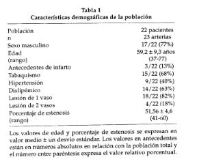
We prospectively included 22 patients and analyzed 23 coronary arteries with intermediate coronary stenoses. The visual estimation of coronary angiogram was determined by two observers in ablind fashion and they were asked to classify thestenoses as significant or not significant with noacknowledgment of other results. Minimal lumendiameter, length and percentage of stenoses weremeasured by quantitative angiography. During thecatheterization there was determined the coronaryflow reserve by Doppler wire measurement classifying the arteries as normal with coronary flow reserve >_ 2 or abnormal with coronary flow reserve < 2.The 23 arteries were classified in two groups depending on the results of the tomographic stress perfusion imaging that were realized within 14 days of the catheterization, groupA (n = 14) as not ischemic and group B(n = 9) as ischemic (if the stress perfusion defect was present in the territory of the intermediate lesion). There were no significant differences between groups A and B with regard to age, coronary risk factors and type of lesions by ACC angiographic classification (A,B or C types). Sensibility, specificity, positive and negative predictive values of the coronary flow reserve results and the visual estimation were determined using the perfusion imaging studies as the gold standard.Correlation between coronary flow reserve and per-fusion imaging studies were analyzed using Kappa and Mc Nemar for paired data.
Results
The quantitative angiography did not show any significant difference in minimal luminal diameter (1.52 ± 0.3 mm versus 1.47 ± 0.3 mm), length (8.04±5.5mm versus 7.97±2.9 mm) and percentage of stenoses(52.4 ± 3.8% versus 49.8 ± 5.5%) between group A and group B, respectively. Sensibility (coronary flow reserve 89% versus visual estimation of coronary angiogram 56%), specificity (coronary flow reserve 93% versus visual estimation of coronary angiogram 43%), positive (coronary flow re-serve 89% versus visual estimation of coronary angiogram 44%) and negative predictive values (coronary flow reserve 93% versus visual estimation of coronary angiogram 57%) of the coronary flow re-serve results were significantly higher than the visual estimations of coronary angiogram. Correlation between coronary flow reserve and perfusion imaging studies was obtained in 91% of cases with an r= 0.81,p<0.0001, confirmed by Mc Nemar test.
Conclusions
This study showed the limited role of the objective or subjective angiographic measurements in the physiological assessment of intermediate coronary stenoses. It was evidenced the great utility of the intracoronary Doppler flow wire in the functional measurement of these kind of lesions that was demonstrated by the high correlation with the perfusion imaging studies.
Downloads
Published
Issue
Section
License
Copyright (c) 2026 Argentine Journal of Cardiology

This work is licensed under a Creative Commons Attribution-NonCommercial-ShareAlike 4.0 International License.